9th European Congress of Thermology, Krakow,
�Poland, May 29 to June 1, 2003:
Abstracts
Comparison of� Infrared Thermography and scanning laser Doppler flowmetry for assessment of the digital circulation in patients with Raynaud�s phenomenon
Al-Awami M, Maca Th, Bartok A, Gschwandtner ME, Minar E
Department of Medical Angiology, University of Vienna, Austria.
Objective: In primary and secondary Raynaud�sphenomenon (RP), measurement of activity or severity, or both, of the digital vascular disease a major challenge. None of the various physiological measurement techniques used in the assessment of patients with primary or secondary Raynaud�sare ideal. Infrared thermography and Laser Doppler blood flow monitoring are non-contact, non-invasive techniques mostly used in the measurement of cutaneous microcirculatory flow� to assess the response to treatment. The objective of this study� was to compare both of these techniques in respect to the severity and activity of RP.
Patients and Methods: Patients suffering from primary or secondary RP were enrolled in this study. The number of� daily attacks of RP and its severity as measured by visual analogue scale on which 0 represented no attacksand 10 the most severe attack ever experienced, were assessed during 8 weeks.� In a temperature and humidity controlled laboratory, a dynamic testing of the digital microcirculation in response to a� standard warm cold challenge test by mean of infrared thermography and laser Doppler perfusion imaging� was simultaneously performed in tow occasions at the following intervals: a) basal� measurement after being adapted to room temperature for� 20 minutes, b) immediately after 1 minute warm challenge (immersion of gloved hands in water at 39�C) , and c) measurements immediately after 1 minute cold challenge (immersion of gloved hands in water at 20�C), d) tow� measurements and 20 minutes later.
Results: Results of the just started study will be given during the congress.
Thermographic assessment of low Ievel laser therapy for treatment of Raynaud�s phenomenon.
Al-Awami M, Schillinger M, Maca Th, Herberg K, Pollanz S,� Minar E
Department of Medical Angiology, University of Vienna, Austria.
Objective�We recently performed a pilot study which suggested that clinical and
thermographic improvements occurred in patients with primary and
secondary Raynaud�s phenomenon (RP) following treatment with low level laser.
In view of these findings, we have proceeded with a double blind,
placebo-controlled study.
Methods�Forty-seven patients suffering from primary or secondary
Raynauds phenomenon were randomly assigned in a double-blind manner to receive
either 10 sessions of low level laser (LLL) distant irradiation (16 f, m,
median age 45 years) or placebo irradiation (21 f, 2 m, median age 46 years)
during winter months. Subjective symptom scores, such as daily frequency and
severity of attacks as measured by a coloured visual analogue scale (VAS) with
0 representing
minimum and 10 representing maximum were assessed. Response to cold challenge
test before and after LLL or placebo treatment was assessed by infrared
thermography. Results- A significant reduction of the frequency as well
as the severity of
RP in patients with either LLL (frequency p<0.0001, severity p<0.000 1)
or placebo treatment (frequency p<0.000 1, severity p=0.02) was found. but
patients in the LLL group exhibited a statistically more significant
improvement at 6 weeks and 3 months of the frequency (p=0.007, p=0.02) and the
severity (p=0.02, p=0.04) of RP. Thermographic response to cold challenge
improved only in patients treated with LLL but not in those treated with
placebo.
Conclusion: LLL treatment significantly lowers the frequency and severity of Raynaud�s attacks in patients with primary and secondary RP- Since this therapeutic modality is a safe, and non-invasive treatment, it might be considered as an alternative to existing therapeutic regimes.
Unusual manifestation of Raynaud�s phenomenon
Al-Awami, Maca Th, Gschwandtner ME,� Maric S,� Minar E
Department of Medical Angiology, University of Vienna, Austria.
Introduction: Raynaud�s phenomenon is a common clinical disorder consisting of recurrent, long-lasting, and episodic vasospasm of the fingers and toes often associated with exposure to cold. The classical progression consists of triphasic colour changes: well-demarcated pallor of the digits leading to cyanosis, pain, and numbness, and followed by a red flush upon rewarming. However, typical episodes involve pallor followed by rubor, with cyanosis present only in severe disease. Other sites, including the tongue, nose, ears, and nipples, can also be affected.
Case report: Here� we report� a patient with unremarkable past medical and surgical history who presented with 10 years history of cold sensitive scrotum with typical Raynaud�s pheno- menon. Through medical and urological exam including Valsalva manoeuvre and colour Duplex scan revealed no pathological finding such as varicocele. Laboratory examinations including auto-antibodies and capillary microscopy were normal. The diagnosis of RP is a clinical one. We could repeatedly objectify it by infrared thermography. To our knowledge this seems to be the first case of such manifestation of RP ever reported
Subject preparation and thermal acclimatization prior to mild cold challenge testing using dynamic thermal imaging
Allen J1, Young AL3, Griffiths B 2, KumarN2,� Murray A1.
Freeman Hospital
Regional Medical Physics Department1
and Department of Rheumatology2,
Newcastle University Department of Physics3. Newcastle upon Tyne. UK.
The mild cold challenge test is frequently used to assess the hands of patients with Raynaud�s phenomonen. Great emphasis is placed on the degree of mild cold challenge, temperature measurement technique, follow-up period, and subsequent analysis. However, protocols involving subject preparation, the key starting point to the measurement process, are also very important. Subjects need to achieve cardiovascular and thermal acclimatization prior to the cold challenge. Typically, 20 minutes is often used for microvascular measurements, irrespective of external ambient temperatures, dress, or subject preparation. The aims of this study were to a) investigate an appropriate pre-test subject preparation protocol for mild cold challenge testing, and to b) assess an appropriate time for thermal acclimatization in normal healthy subjects.
The pre-test preparation protocol was compiled from information obtained from five European microvascular measurement centres who undertake mild cold challenge testing of the hands. The protocol asked subjects to follow guidance on diet and medication, dress, relaxation, and hand preparation within specified times prior to their study. Initially, subjects completed a health questionnaire to exclude cardiovascular disease, persistently cold hands or Raynaud�s phenomenon. The minimum time for thermal acclimatization was then estimated from hand temperature measurements from 16 normal subjects (8 male and 8 female) of age 33“12 years (mean”standard deviation). All subjects gave their written informed con- sent. Each subject followed the pre-test preparation protocol before sitting in a cool temperature-controlled room for 20 minutes (local study temperature 17“1 oC). This was sufficient to result in peripheral vasoconstriction but without inducing shivering or significant discomfort. Each subject then sat quietly in a medical infrared imaging facility (ambient temperature 24”1 oC) for 40 minutes whilst their hand skin temperatures were measured at 1 minute intervals (FLIR SC300 thermal imaging system). The operator and subject were blinded from the measurements during this follow-up. The temperature data were processed using dedicated FLIR ThermaCam Researcher image processing software, with skin emissivity assumed to be 0.97. Each sequence of images was studied twice and averaged to give an estimate of the time taken for the hands to reach a plateau with warm and evenly distributed temperatures.
The median (2.5-97.5 percentile) time for the 13 subjects (6 male and 7 female) whose hands re-warmed within the follow-up period was 14 (9-31) minutes. The recovery generally showed bilateral similarity between the right and left hands and there was no significant difference between males and females (Mann-Whitney test). When all subjects were considered, including the 3 that did not adequately re-warm, the median time increased to 18 minutes, with no significant difference between the sexes. Four of the 16 normal subjects (25%) had not recovered within 30 minutes.
The pre-test protocol was acceptable for the subjects. We have shown that 30 minutes is not always long enough for acclimatization, even in normal subjects. These preliminary findings have implications for cold challenge testing of the hands using dynamic thermal imaging.��
Thermal Features Of Hot Packs
Ammer K, Melnizky P
Ludwig Boltzmann Research Institute for Physical Diagnostics,� Vienna, Austria
The temperature course of two self-heating and three passively heated packs was measured by infrared thermal imaging. Measurements were performed at a room temperature of 24�C. The packs were put on the ground with a woollen blanket underneath to prevent heat loss by conduction.
The size of the packs, the maximum temperature and duration of heat dissipated from them was variable. The highest temperature values were observed in self-heating packs. One self- heating pack reached a maximum temperature of 55�C, but decreased in mean temperature by 12 degrees within 20 minutes. The other self-heating pack reached a maximum temperature� of 40 degrees, but stayed at a mean temperature of 33.5�C for at least three hours. With parafango hot packs the temperature fell from 43 �C to 33�C within 20 minutes. Mud packs presented with a similar cooling course. A newly designed re-usable pack showed different peak temperatures depending on the temperature of the storage case. When stored at temperatures of 55 or 70� degrees the peak mean temperature was 38 and 43 degrees respectively. Independently from the starting value, the mean temperature of this pack decreased by 10 degrees within 20 minutes.
The different materials used for therapeutic hot packs affect the course of temperature change and may therefore have different heating effects on the skin during heat treatment.
An Introduction Into Thermal Physiology
Ammer K.
Ludwig Boltzmann Research Institute for Physical Diagnostics, Vienna, Austria
Thermal physiology describes all body functions related to thermal energy given to or removed from a living body. The most important physiological system in this context is temperature regulation, which keeps the temperature of the inside of the body on a constant level. This is achieved by changing the temperature in the outside of the body varying the superficial blood flow and heat production or activation of additional cooling mechanisms such as evaporation of sweat on the skin surface. The human body uses sympathetic nerve fibres for information spread related to temperature regulation. However, temperature regulation is only one function of the autonomic nerve system. Its main function is the non-voluntary control of smooth muscle fibres.
Strong interactions exist between temperature regulation and the cardiovascular system, also with fluid and energy control Heat generated by contraction of striated muscle fibres is the most important internal heat source of the body. Understanding the mechanisms of heat exchange of the body with the environment is essential for correct interpretation of temperature patterns on the body�s surface. Any disturbance of the heat balance of the body is followed by temperature regulation, which keeps the deep body temperature close to the set point. Exhausting the regulation capacity of the system leads to a new set-point i. e. either increase (hyperthermia) or decrease (hypothermia) of the core temperature. The mean skin temperature and the core temperature jointly determine the regulation process. Skin temperature is the result of the heat storage of the body and the thermal environment. The law of physics for heat transfer provides the means of predicting the mean skin temperature under defined conditions.
Various mechanisms unrelated to temperature regulation may affect the diameter of superficial skin vessels, resulting in different levels of skin temperature. Temperatures on the surface can only be correctly interpreted if the condition of the thermal environment is known. It is not true to assume that the surface temperature is synonymous with perfusion or that blood flow is exactly the same as surface temperature. However, very specific responses of vessel control do occur in certain thermal conditions.
Temperature regulation under working conditions is of practical importance to man, especially for research into safety procedures in extreme temperature conditions. The balance be- tween protection against either heat or cold and gross endogenous heat production can be a very difficult challenge. In such a situation interactions of temperature regulation with the cardiovascular system and fluid balance become significant.
Many physiological functions are related with the thermal phenomenon, but not all are the result of temperature regulation. Basic knowledge of thermal physiology is necessary for the correct interpretation of human body temperature measurements.
Skin Temperature After Intake Of Sparkling Wine, Still Wine And Sparkling Water
Ammer K,.Melnizky P, Rathkolb O.
Ludwig Boltzmann Research Institute for Physical Diagnostics, Vienna, Austria
An increase in skin temperature after intake of 8ml alcohol has recently been reported. It is generally believed that fast intake of sparkling wine is associated with facial flush. We investigated� the effects of drinking sparkling wine and whether this can lead to higher skin temperature compared to drinking a glass of non-sparkling wine or sparkling water.
8 women and 4 men were included in the study. All subjects acclimatised with bare arms and legs in a room temperature of 24�C. After acclimatisation thermal images of the face, both hands (dorsal view) and both knees (anterior view) were taken. Thereafter a bolus of either 70 ml sparkling wine (11,5% alcohol) or 70ml� still wine (11,5% alcohol) or 70ml sparkling water was given. Another series of images was performed 15 minutes later. Mean temperatures of face, hands and knees were determined and statistically analysed.
An increase of skin temperature in all investigated body regions was observed after alcohol intake. No significant difference in temperature elevation was found between sparkling wine and wine without gas. An increase of skin temperature was not detected after drinking sparkling water.
The increase of skin temperature after alcohol intake was confirmed. However, the presence of CO2 in sparkling wine seems to have no additional influence on skin temperature.
Standard Positions For Imaging The Human Body With Infrared
Ammer K
Ludwig Boltzmann Research Institute for Physical Diagnostics, Vienna, Austria
A protocol for capturing a series of infrared images that cover the whole human body was developed at the Thermal Physiology Laboratory, School of Computing, and University of Glamorgan in UK. A total of 24 views were specified including 3 views of the whole body.
The consistency of positioning of the standard views �Face�, �Dorsal Neck�, �Upper Back�, �Anterior Left Arm�, �Dorsal Hands�, �Both Knees Anterior� �Lateral Right Leg� and �Plantar Feet� was evaluated. The distance, measured in pixels, from the upper, lower or side edge of the image to anatomical landmarks was used for evaluation. The cross section analysis tool of CTHERM software was used for the determination of distances.
The highest variation in positioning was found in the hands and feet. The face varied in a very narrow range. Table 1 shows the variations in positioning of all the investigated views.
The repeatability of standard views varied according to the body regions investigated. Individual dimensions of these body regions contribute to the variation of positioning. In the case of dorsal hands the distance between both little fingers may be longer than the distance from the wrist to the tip of the middle finger. Such a condition prevents the precise positioning in a defined manner. Similar conditions may occur in the views Upper Back, and Anterior Knees. According to the results of this investigation the rules for positioning and image capture of dorsal hands, upper back and anterior knees have been modified.
�
|
Table 1 Variations in positions
|
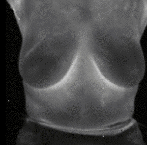
Infrared-Thermography of the Upper Extremities� of Breast Cancer Patients
Bartok A, Maca Th,Berger A, Al-Awami, M, Herberg K, Minar E.
Department of Medical Angiology, University of Vienna, Austria.����������
Background: Infrared-thermography (IRT) has been widely used as a screening method for breast cancer, which did not succeed in reaching the best sensitivity. IRT might be helpful in detecting first hints for secondary arm lymphedema.
Patients and methods: We tested 12 women (mean age 56,4 years) with breast cancer the day before, 1 week and 6 months after axillary lymphadenectomy of the upper extremities by IRT.
Results: We observed a clear trend towards a higher temperature on the affected upper arm (treated vs. untreated arm: 32,4 vs. 31,7�C) and in the axilla (treated vs. untreated arm: 32,4 vs. 33,7�C) compared to the opposite side at baseline. This difference proved to be markedly diminished on the forearm (treated vs. untreated arm: 31,9 vs. 31,3�C). One week past operation, we could detect a generalised temperature rise on both upper- (treated vs. untreated arm: 34,3 vs. 33,6�C) and forearms (treated vs. untreated arm: 33,4 vs. 33,1�C) and in the axilla (treated vs. untreated arm: 35,9 vs. 35,2�C), followed by a drop of temperature on the upperarm (treated vs. untreated arm: 32,8 vs. 32,7�C), on the forearm (treated vs. untreated arm: 31,9 vs. 31,8�C) and in the axilla (treated vs. untreated arm: 34,6 vs. 34,3�C) after 6 months. These primary results did not reach statistical significance, probably due to the small number of patients. But arms without lymphoedema (30,6�C) presented with significantly less mean temperature than arms with moderate (31,2�C) or high-grade lymphedema (32,9�C).
Conclusions: IRT might be a useful tool for early detection of candidates for secondary arm lymphoedema.
Results of thermographic assessment of periodontal tissues in children suffering from decompensated IDDM versus the time of the disease
D.Burchardt
Department of Paediatric Dentistry,Institute of Dentistry, K.Marcinkowski , University of Medical Sciences, Poznań, Poland
An important aspect of the health care of the patients in develop- mental age suffering from Insulin Dependent Diabetes Mellitus� (IDDM) is� the prevention of early and late complications through the control of metabolic compensation. Changes of microangiopathic and macroangipathic character are most often a consequence of a long time hyper- glykemia. However, a relation between the time of the disease and the occurrence of angiopathies is still a subject of discussion.
The aim of the study was assessment of the response of periodontal blood vessels to a cooling stimulus in patients in developmental age suffering from decompensated IDDM.
�The subjects of the study were 32 boys and girls aged 10-19 (mean age 16.0) suffering from IDDM. The subjects were allocated� into two groups those suffering for 4-9 years (73.8%) and those whose disease lasted for 10-14 years (26.2%). High values of HbA1 (mean 11.2%) indicated metabolic decom- pensation. The oral cavity hygiene status was assessed according to the Pl-I index according to Silness & L�e, while the periodontal tissue status was described with GI according to Silness & L�e. In the thermographic assessment we analysed the mean weighted values of temperature measured before the stimulus (T0) and 1 minute (T1), 2 minutes (T2), 3 minutes (T3) and 4 minutes (T4) after the stimulus.
�In the group with shorter lasting disease the values of Pl-I varied from 0.0 to 3.1(median 0.7), while in the group with longer lasting disease these values varied from 0.1 to 2.2 (median 0.6). According to the results of non-parameteric Mann- Whitney test, no statistically significant differences were noted (p>0.05). The hygiene status was satisfactory and comparable in both groups. No changes in periodontal tissues were found in 2.2% of the children suffering for 4-9 years and in 6.3% of those treated for 10-14 years. In the whole group studied the mild gingivitis was observed in 77.8% of the children with shorter lasting disease and in 62.5% of the children with longer lasting diseases (over 10 years). Medium stage peri- odontitis occurred in 17.8% of the children with shorter lasting disease and in 31.2% of those with longer lasting disease. The acute stage of periodontitis was noted only in 2.2% patients with shorter history of treatment. The results obtained were analysed by the nonparameteric Mann-Whitney test which did not reveal statistically significant changes between the groups (p>0,05). The mean weighted temperatures in the group of the children with longer lasting disease were: T0=33.6�C, T1=27.0�C, T2=29.0�C, T3=30.1�C, T4=31.2�C, while in the group with longer lasting disease: T0=32.8�C, T1=26.4�C, T2=27.2�C, T3=27.9�C, T4=28.8�C.
�The results obtained indicate that the method of thermography can be an important supplement of clinical diagnostic methods used to assess the periodontal status in IDDM sufferers.
Digital Infrared Thermographic Imaging of Osteoporotic Compression Fracture in Elderly Patients
Cho YE. Kim YS
Yongdong Severance Hospital, YonseI University College of Medicine; Seoul, Korea
Digital Imaging Thermographic Imaging is a diagnostic tool of painful conditions in neuromuscular skeletal disease. lt shows high sensitivity in pain detection and correlation with the clinical condition pre- and postoperatively.
We applied digital infrared thermal imaging (DITI) for osteo- porotic compression fracture in elderly patients having multiple compression fractures to evaluate the efficacy of DITI to differentiate between a new lesion and old compression and to correlate DITI with postoperative clinical result. Clinically it is difficult to differentiate the symptomatic lesion in multiple compression fractures in elderly patients.
78 patients operated by vertebroplasty due to compression fracture were included. They were investigated by plain X-rays, MRI, bone scan and thermal imaging. Thermal changes were analyzed by the anatomical location and the thermal difference preoperatively. Thermal images were correlated in anatomical location with MRI and bone scan and eith pain severity measured by VAS.
73 of 78 patients showed a marked hyperthermia on the site of the lesion (93.6%). The hyperthermic lesion was well correlated with symptomatic new lesions. The thermal change was stable on thermal imaging for 6 months after trauma.
After vertebroplasty, the pain was reduced in all patients and the thermal difference was smaller than in preoperative images in 63.1%.
DITI is very effective to detect the pain due to compression fracture and has a high diagnostic potential efficacy to differentiate new lesion from old lesion. Temperature changes correlated well with the postoperative clinical results.Thermal Imaging is very useful for the diagnosis and postoperative follow up of osteoporotic compression fractures.
Teletermographic� Images Of Brain Lesions (Tumours Or Vascular Diseases )
Dalla Volta G.
U.O.Neurologia �Istituto Clinico Citta� di Brescia �Italy
The autonomic nervous system (ANS) plays a key role in the regulation of the vascular resistance thrhough a direct influence on skin microcircle. Vascular tone is the end result of the combined action of the sympathitic system (vasoconstrictor) and the parasympathetic system (vasodilator) on vascular smooth� muscle cells, with a minor role played by several circulating molecules with a vasoactive effect (i.e adrenalin, vasopressin, et al). As far as the facial district is concerned, any structural or functional injury on the trigeminal system may result� in the antidromic release of vasoactive substances such as CGRP, P substance (???), NO, VIP etc.� Any modification of such equilibrium can be reliably detected as an abnormal pattern of skin temperature by a thermographic investigation. Given the functional interaction of the peripheral nervous system with different cortical and subcortical areas, as well as with the endocrine system through the hypothalamus-pituitary axis, any structural abnormality of the human brain (such as a neoplastic or a vascular lesion) may determine a functional impairment of the autonomic nervous system, leading to a modification of the thermographic pattern. A hypothermic pattern is the consequence of vasoconstriction due to an sympathetic hyperactivity while a hyperthermic pattern is the result of an uncontrolled vasodilation due to the over-expression of the parasympatethic system. The aim of the current study is to support this evidence with a series of thermographic pictures from patients with carotid aneurysms, lesions of the cavernous sinus, cerebral tumors and cerebral infarct.�
Database of Standardized Thermal� Signatures of the Breast Upgrade
Diakides NA, Diakides M
Advanced Concepts Analysis, Inc., Falls Church, VA
This program is a collaborative effort which will develop a web-based database of breast thermal images aimed at characterizing and quantifying breast signatures of� both tumors and benign conditions.� It is a new initiative sponsored by the Deputy Assistant Secretary of the Army for Installations and Environment (Environmental Safety and Occupational Health).� The coalition consists of two medical research centers, Ville Marie Medical and Women�s Health, Canada, and Elliott, Haley and Head Breast Cancer and Treatment Center, USA, the Air Force Virtual Distributed Laboratory, AFRL, George Washington University and Advanced Concepts Analysis, Inc.� The strategy, objectives and goals of this program will be discussed as well as the methodologies.�� Uploading and downloading of� radiometric images by multiple medical centers to a� secure central� website will be presented . Need for a common image format will be discussed.�� Analysis of preliminary results obtained from six hundred patient data will be addressed.
Applications of Thermal Imaging in Human and Veterinary Medicine
Fik�ckov� H 1, Meszaro�ov� M 2, Jirman R 3, Navr�til L 1,4, Z�vi�ek M 5, Drastich A 5, Dub P5
1 Charles
University Prague,1st Medical Faculty, Institute of Biophysics and Informatics,
CR
2 Charles University Prague, Dept.of Anatomy and
Biomechanics FTVS UK,CR
3 Charles University Prague,1st Medical
Faculty, Department of Stomatology, CR
4 University of South Bohemia, Cesk�
Bud�jovice, Faculty of the Health and Social Studies, Department of Radiology,
CR
5 Brno University of Technology,
Department of Biomedical Engineering, Brno, CR
Besides the study concerning breast cancer detection, which is presented in another contribution, we have tried several other applications of thermography in human and veterinary medicine at our department. This article presents short overview of these applications.
The first study should find the thermographycal changes related to overuse tendon injuries of race horses. This disease is supposed to be an� inflammation process and the term tendinitis is used, although etiology remains unknown. Each inflammation causes local increase of temperature and should be thermographically visible on the overlaying skin surface. Some studies made mainly in United States say that there is a correlation between temperatures changes and tendon injuries; however, our research shows that temperature changes are only rarely present in this injury and thermography is of little use to evaluate or detect it. We thing this finding also means that inflammation is generally not present and this disease should be termed tendinosis.
Topic of the second study should find out if thermography could be used as a diagnostic tool to distinguish patients with arthralgia of the temporomandibular joint (TMJ) from healthy subjects. Temporomandibular joint disorders (TMD) have been identified as a major cause of non dental pain in the orofacial region and are considered to be a subclassification of musculoskeletal disorders. Although current imaging techniques (e.g. radiography, arthroscopy, X-ray CT or MRI) can image both soft and hard tissue, there was reported a poor correlation between signs and symptom of TMD and MR imaging and radiographs. There is an absence of diagnostic method, which can objectively assess severity of clinical sings of TMD and therefore determine the need for treatment and evaluate the treatment efficiency. The aim of this study is to evaluate the diagnostic potential of the infrared thermography for diagnostics of patients with unilateral arthralgia of TMJ of arthrogenous origin. The first results will be presented on conference since they are not available at the time of submission of this abstract.
Raynaud�s Phenomenon in Chronic Fatigue Syndrome.
Harding JR,� Llewellyn M.
Royal Gwent & St Woolos Hospitals, Newport, Gwent, UK
A previous case report showed dramatic response of severe Raynaud�s phenomenon in a young girl with Chronic Fatigue Syndrome to treatment with selective serotonin re-uptake inhibitors (SSRI�s).�
Patients with Chronic Fatigue Syndrome with signs and symptoms of Raynaud�s phenomenon were assessed by infra- red imaging of the hands and wrists before and after a cold challenge, with quantification and analysis.� This was per- formed prior to treatment with SSRI�s and repeated after 4-5 weeks treatment in a series of patients in an on-going study.�
A dramatic reversal from severe thermologically proven Raynaud�s phenomenon initially, to complete normality following treatment was observed.� This is interesting as the only treatment given was with an SSRI anti-depressant, no treatment with vasodilators being given
Physiological Factors affecting the Reliability of Infrared Thermometry: Choice of Ear
Heusch AI ,McCarthy PW.
Welsh Institute of Chiropractic,� School of Applied Sciences,� University of Glamorgan;� Pontypridd, Wales (UK
Infrared tympanic thermometry (ITT) is the preferred option for medically assessing body core temperature, but there has been increasing concern over its reliability (McCarthy et al., 2002).� Earlier studies have reported differences between the left and right ear temperatures in the same person (Joly et al., 2001; Modell et al., 1998).� We have studied this further by recording temperature bilaterally and noting which ear was recorded first.� We investigated three groups of university stu- dents; Group 1, assessing the right ear first then the left; Group 2, assessing the left ear first and then the right and Group 3, randomly choosing an ear to be first.
|
First ear recorded from |
Right (n=15) |
Left (n=40) |
Random (n=13) |
Total (n=68) |
|
First ear temp. Mean�1SD |
36.3�0.6 |
36.8�0.5 |
36.5�0.4 |
36.6�0.5 |
|
Second ear temp.Mean�1SD |
36.5�0.6 |
36.9�0.6 |
36.6�0.4 |
36.8�0.6 |
|
Wilcoxon (p=) |
0.019 |
0.009 |
0.017 |
<0.001 |
|
Parity/regression intercept temp |
37.4 |
39.7 |
38.1 |
38.0 |
|
Spearman(r=) |
0.682 |
0.810 |
0.884 |
0.849 |
The results indicate that current medical practice of taking only one ear temperature might lead to assessment error. This could significantly affect the temperature recorded in the patient records and thus potentially affect the treatment protocol adapted.� It is suggested that clinicians consider taking auricular temperature bilaterally and then choose the highest reading as being the best estimate of �core� temperature.
References
Joly LM, Giraudeau B., Monchi M,� Oswald, AM. [Is measurement of body temperature by infrared tympanic thermometry re- productible?] Annales Francaises d�Anesthesie et de Reanimation 2001; 20(10): 833-7
McCarthy PW,� Heusch AI,� Kenkre J, Machin G, Suresh J. Letter to the editor.� The Lancet2002; 360(9348): 1882-3
Modell JG, Katholi CR,� Kumaramangalam SM,� Hudson EC, Graham, D.� Unreliability of the infrared tympanic thermometer in clinical practice: a comparative study with oral mercury and oral electronic thermometers.� Southern Medical Journal 1998; 91(7): 649-54
The Cold Challenge Test and Infrared Thermography for the Objective Assessment of Digital Vasospasm In Out-patients Referred to A Dedicated Raynaud�s Phenomenon Clinic
Howell KJ, *Smith RE, Wilson H, Black CM
Department of Rheumatology and *Department of Medical Electronics, Royal Free Hospital, London, UK
Local patients referred to the Rheumatology Department of the Royal Free Hospital for suspected primary Raynaud�s phenomenon are asked to attend a Testing Clinic prior to assessment by a physician or specialist nurse some weeks later. The Testing Clinic protocol comprises nailfold capillaroscopy, autoantibody blood screening and infrared thermography to assess the rewarming rate of the hands after their cold challenge in water at 15�C for one minute.
In January 2003 we reviewed thermographic data from 84 patients referred consecutively to the Testing Clinic between the date of acquisition of our FLIR SC500 Thermacam FPA imager in April 2001, and December 2002. 74 patients attended for subsequent clinical assessment, when the Testing Clinic results were available to the clinician. 57 patients had a diagnosis of autoantibody-negative primary Raynaud�s phenomenon (RP) confirmed by the examining physician. (45F: mean age 39.5 yrs, SD 14.5 yrs. 12M: mean age 31.7 yrs, SD 15.4 yrs). Seven further patients were autoantibody positive, APRP (all F: mean age 45.6 yrs, SD 12.8 yrs). The physician diagnosed three of the remaining patients with a connective tisssue disease (2 LcSSc, 1 UCTD, all F). Seven other patients were considered negative for a diagnosis of Raynaud�s phenomenon or CTD and either referred forward to alternative clinics or discharged (3M, 4F). Only 2 patients tested had abnormal nailfold capillaries (1 LcSSc, 1 RP).
We took a mean percentage finger temperature recovery of >90% and a baseline mean finger temperature of >30�C as cut-off points for normality. Of the 57 patients diagnosed with RP, 11 were deemed normal by our thermographic criteria. 42 RP patients had an abnormal dynamic response (7 of whom had a baseline mean finger temperature above 30�C), and 4 had a normal dynamic response to cold challenge but an abnormal baseline temperature (range 26.3�C � 28.9�C). Of the 7 APRP patients, 3 were normal according to our thermographic criteria.
We conclude:Further analysis is required to improve the discriminating power of the thermographic assessment
Consideration of both baseline mean finger temperature and dynamic response are required for adequate assessment of an individual patient.
Symmetry in classification of healthy and malignant breasts using thermography
Jakubowska T, Więcek B.,. Wysocki M, Peszyński-Drews C.
Technical University of Lodz, Laser Therapy Center & Institute of Electronics
One of the principal feature of breast thermographs is symmetry of temperature pattern. Thermal images of breast are usually asymmetrical in pathological cases. The aim of this re- search is to find image parameters that describe the symmetry of breast thermographs and to use this parameters to separate thermal images of healthy breast from malignant ones.
The thermographs of 32 healthy patients and 10 patiens with malignant tumour were analysed� Four images were recorded from every patient which represented each breast in a perpendicular and lateral view. Histograms were created for these images and on the basis of that we calculated statistical para- meters such as� mean temperature, standard deviation, variance, skewness and kurtosis. The� absolute differences of parameters� between left and right breast were also determined. The degree of symmetry was based of these differences.
Mean temperature in healthy group was equal to 30.24 � 1.78 in the perpendicular view� and 29.75 � 1.86 in the lateral view.The mean temperatures were higher in the malignant group than in healthy subjeczs� 8 of 10 cases. Furthermore, there were 6 cases out of 32 in the healthy group whose mean temperature exceeded the normal temperature range. Therefore, it� is necessary to analyse symmetry. The comparison of mean temperature did not� sufficiently separate pathological findings from healthy breasts. Among the analysed parameters , skewness was the most useful parameter for the classification of the images. In the healthy group, the absolute differences of skewness was equal to 0.41 �0.34 in the perpendicular� position and 0.63 � 0.46 in the lateral view. At least in one view of the group with pathologies, this difference was higher than in healthy controls.
|
|
|
|
Fig.1: Thermal breast image with malignant tumor (on left side) |
Fig.2: Thermal image of healthy breast |
�
Content Based Image Retrieval a database of medical thermal images.
Jones BF
School of Computing and Technology, University of Derby, Derby,� UK
Content-based image retrieval (CBIR) [1] aims to identify images according to their subject rather than a textual description or title.� The images are chosen according to parameters derived from their pixel values, colour, texture and so on.� The design of such a system demands that
1.image features for identification must be specified;
2.suitable algorithms to capture the chosen features must be developed;
3.an index and appropriate data structures must be formed;
4.a user-interface for making queries must be designed.
CBIR systems [2] for visible images are based on pixel-level features (e.g. colour), global features (e.g. histograms), textural features, object features (e,g, Hough transform) and conceptual features (e.g. time, location) [1].
A database of thermal images of normal subjects is under construction at Glamorgan University; this will provide a resource of information for researchers and clinicians who use thermal images as a diagnostic tool.� The aim of this project is to investigate the use of CBIR techniques to retrieve images from a database of medical, thermal images which have similar cha- racteristics to a given image that needs to be interpreted.� The associated clinical findings of retrieved images may provide pointers to clinicians of underlying problems in the given image and may help to develop hitherto unsuspected insights into manifestations of problems for the researcher.
The use of moments may provide suitable image features for a CBIR system that could be developed for retrieving thermal images [3].� Although moments are normally used with binary images, they may also be used with grey levels. The moments are normalised to be invariant with respect to translation, rotation and scaling.� Moments express the degree of symmetry within the image and measure skewed distributions of pixel levels.� It is well known [4] that asymmetrical temperature distributions and hot and cold spots are strong indicators of an underlying dysfunction.
References
[1] Gong, Y, Intelligent image databases: Towards advanced image retrieval, Boston:Kluwer Academic Publishers, 1998, ISBN 0-7923-8015-0.
[2] Ogle, V E, Stonebraker, M, Chabot:Retrieval from
a relational database of images, IEEE Computer, 28, 1995.
[3] Schalkoff, R J, Digital image
processing and computer vision, New York:Wiley, 1989, ISBN 0-471-50536-6.
[4] Uematsu, S., 1986 , Symmetry of skin
temperature comparing one side of the body to the other, Thermology,� 1, 4-7
An Update to the Searchable Archive of Thermal Imaging for Medicine Papers
Jones BF 1, Ring EF �,� Jones CD �
1School of Computing and Technology, University of Derby, Derby, UK
�University of Glamorgan, School of Computing,�
Pontypridd, UK
Infrared thermal imaging has been applied in medical research since 1960 in several research centres in Europe, the USA and Japan. Many papers were published in the journals ACTA THERMOGRAPHICA and THERMOLOGY. During this period, several important basic principles of thermal imaging were established, such as the thermal symmetry inherent in a healthy subject. Both these journals ceased publication some years ago and this important early research has become difficult to access. In 2000, with funding from The US National Technology Transfer Center, Bryan Jones and Francis Ring produced �A Searchable Archive of Thermal Imaging for Medicine Papers on CD-Rom�.� This made the material widely available to researchers in medical thermal imaging. Here we present an update to the archive that includes Thermology International abstracts from 2001 to 2002. The updated archive also includes a searchable bibliography of papers published on thermology or temperature measurement between 1989 and 2002, contributed by Kurt Ammer.
Reference
Jones B.F, Ring E.F. A Searchable Archive of Thermal Imaging for Medicine Papers on CD-Rom. Thermology International. 10(4): 201-203, 2000.
Optimised registration of infrared images for comparison of standard views of normal human subjects
Jones C, Plassman P, Ring EF.
University of Glamorgan, School of Computing, Pontypridd, UK
The CTHERM image capture package provides overlays with outlines of the standard views, however, individual images are never precisely aligned and human subjects are not the same shape. Consequently the images need to be �registered�. This process is divided into a linear stage (rotation, translation and scale) and a non-linear warping stage which takes account the difference in individual body shape.
To identify the most suitable method for the non-linear registration stage, we compare two appropriate warping algorithms; The Thin-Plate Spline algorithm (Bookstein 1989) and a Feature-Based base algorithm (Beier & Neely 1992).
Results indicate a combination of linear registration (to account for changes in view alignment and camera position) and warping using thin-plate splines (to account for differences in body shape) is optimal for minimising variability of standard views of normal subjects.
References
T. Beier and S. Neely. Feature-Based Image Metamorphosis. In Computer Graphics, 26(2): 35-42, New York, NY, July, 1992. ACM SIGGRAPH. Proceedings of SIGGRAPH �92.
Bookstein, F.L. 1989. Principal warps: thin-plate splines and the decomposition of deformations. IEEE trans. Pattern Analysis Machine Intelligence. 11(6): 567-585, 1989.
Application of Thermovision in the Diagnosis of Inflammatory Changes of Nasal Sinusitis
Jung A1,� Zuber J1, Kalicki B1,� Murawski P1,� Muszyńska J1 ��Ligęziński A2
1 Thermal Laboratory of Pediatric and Nephrology Clinic of Military Medical Institute,� Warsaw, Poland
2 ENT Department Clinic of Military Medical Institute, Warsaw, Poland
The aim of this study was to image the temperature arrangement in anterior and posterior nares in patients with sinusitis.
The nasal cavity with nasal sinuses is a space for which it is important to keep a constant temperature. Physiological activities of the nose are closely connected with the air-flow through the nose, creating the only physiological respiratory tract. In case of high temperature of the surrounding air, the breathing path through the mouth is used. The same also occurs in cases of anatomical abnormality of the upper respiratory tracts, lung and the circulatory system diseases and during exercise. But it should be emphasized that only breathing through the nose ensures the proper thermal cycle of the air inspired from the environment. Warming of the inspired air occurs due to heat transfer from the blood circulating in the vessels of subcutaneous tissue, mostly in the cavernous plexuses in the posterior nares. The warmed air reaches a temperature of 32-34�C independently of the temperature of the breathed air . The process of warming the inspired air can be investigated in the study of the heat emission registered by the thermal imaging camera . Under physiological conditions it assumes a symmetrical picture in both nostrils with regularly rising temperature from the anterior to posterior nares. There are also two other factors which decide the proper physiological activities of the nasal sinuses, namely ventilation and permeability. Ventilation depends on keeping proper width and permeability of the fissures connecting the sinuses with the nasal cavities. Permeability is affected by the secretion of the mucus and transporting it outside the sinuses. The whole process can be shown by thermo- graphic examination. Under inappropriate conditions the symmetry of the thermographic picture can be disturbed. The standardization and objective measurement of thermal changes has paved the way for large-scale thermal imaging usage in the diagnosis of inflammatory focus). The possibilities of differentiation the etiology of mucous membrane chan- ges of allergic or inflammatory background are of specially interest and value in clinical practice.
The thermographic evaluation of cases with a range of inflammatory changes in the area of the paranasal sinuses was comparable with a radiological image. It also provided non- in- vasive monitoring of the pathological process, without the need for computed tomography in the first case.
Infrared Imaging of Angiomatosis Syndrome (Klippel � Trenaunay Syndrome
Jung A1, ZuberJ1, Kalicki B1, Perdzyński W2, Klewar M3,� Murawski P1
1 Thermal Laboratory of
Pediatric and Nephrology Clinic, Military�
Medical Institute, Warsaw, Poland
2 Department of Children Surgery, Military Medical
Institute, Warsaw, Poland
3 Radiology Department of Military Medical Institute,
Warsaw, Poland
One possible application of thermography in medicine is the imaging of� venous vessels.
�In this paper the authors describe a case of a 15-years old girl attended the Children�s Surgery Clinic, Military School of Medicine in Warsaw with angiomatosis of the right lower limb (Klippel � Trenaunay�s syndrome). Large angiomas of the foot, calf (especially laterally), knee and thigh were assessed.
Doppler ultrasound examination of the right thigh confirmed an extensive venous angioma involving superficial and deep veins. Blood flow in these veins was evaluated in the seated and supine positions. There was no evidence of thrombosis. An oscillating blood circulation was observed during the Valsalva maneuver and during compression. Blood flow with in the angioma was not visualized. Cavernous angiomas were also examined in the foot and on the anterior and lateral surface of the thigh. In the left lower limb the popliteal vein and the long saphenous vein were narrower than the same veins in the left contralateral limb. The blood flow in the deep veins of the right Iower limb was maintained but it was slower than in the Iower left limb. There was normal blood flow in the arteries of both lower limbs. On venography, there was a conglomeration of dilated superficial veins. The deep venous system was not visualised, except for a portion of popliteal vein. At surgery very wide (2-2.5cm) thin-walled angiomas were exposed and separated superficial to the fascia, with many connections between them and between the vessels situated sub-fascially. After ligation, the angiomas, which spread out in the upper calf, were resealed. Post operatively the wound healed well. Six months later there was no pain or inflammation in the calf.
On post-operative Doppler ultrasound there were no angiomas present in the operated area. The visualised blood flow through the popliteal vein and long saphenous vein was faster than before the operation but still slower than on the left side. Thermo- graphic investigation of the right lower limb before the operation showed extensive hyperthermal areas in the right thigh, extending into the right popliteal and calf areas. The hyper- thermal areas corresponded with� the images of conglomerates of dilated veins on Doppler ultrasound. Post operatively, the areas of increased tem�perature were significantly smaller. The temperature gradient between the area of abnormal veins and the surrounding tissue was about 4.5�C pre-operatively, however, after the operation the gradient decreased to 2.5�C. Thermographic visualisation strictly correlated with the clinical assessment of the post-operative state.
Infrared� Thermographic Imaging of Critical Leg Ischaemia
Jung A1,� Zuber J1, Kalicki B1,� Stańczyk M2,� Osiecki M3,� Twarkowski P3,� Murawski P1
1 Thermal Laboratory of Pediatric and Nephrology
Clinic of Military Medical Institute, Warsaw, Poland
2 II Surgery Department of Military Medical Institute,
Warsaw, Poland
3 Radiology Department of Military Medical Institute,
Warsaw, Poland
Infrared Thermography can be used to evaluate and monitor� ischaemiaof the lower limb.�
A case of a 60-year old female patient (G.A.) with 10 years history of arteriosclerosis obliterans with ischaemic right lower limb, lumbar symphatectomy and succesfull functioning of right femoropopliteal prosthesis is presented.
The patient was admitted to the surgical department with symptoms and signs such as pain at rest, muscle stiffnes and distanse of intermittent claudication of 50 m. On physical examination trophic changes of the skin of the right lower limb were observed. Doppler ultrasound and angiography confirmed occlusion of the right femoropopliteal prosthesis. The indication for surgical intervention was limb salvage. During the operation an occluded femoropopliteal prosthesis was found. The surgical procedure included restoring the prosthesis patency by embolectomy and profundoplasty with a venous patch above the proximal part of the deep femoral artery. Because there was no evident clinical improvement of the lower limb perfusion, surgical procedure was followed by 7 days aloprostadil (Prostavasin - PGE1)� administration. Due to aggravation of the right lower limb ischaemia our patient required below knee amputation.
A second case a patient (B.S.) with critical leg ischemia is presented. He was treated with good results by aloprostadil.
�The infrared thermography� findings showed� a strong correlation with� the clinical picture.
Infrared Imaging of Varicocele
Jung A1,� Zuber J1,� Kalicki B1,� Perdzyński W2,� Klewar M3,� Murawski P1
1 Thermal Laboratory of Pediatric and Nephrology Clinic, Military Medical Institute, Warsaw, Poland
2 Department of Children Surgery of� Military Medical Institute, Warsaw, Poland
3 Radiology Department of Military Medical Institute,
Warsaw, Poland
In this study infrared thermography was used to imagine the pre- and postoperative state of varicocele.
A 14-year-old boy (K.K.) with left side varicocele was hospitalised in the Clinic of Children�s Surgery MUSM; varicocele In the Doppler USDoppler USG study a widened seminal vein with considerably slowed blood flow was revealed.
During the operation 7 widened blood vessels were found and 1 cm. long section of veins between the ligatures was resected. The recovery after the operation was satisfactory. In the US investigation made after the operation varicoceles were not found to be present.
In the thermographic study before the operation, a homogenous focus was visualised in the area of the left spermatic cord and the area of the left groin. The temperature gradient between the hyperthermal focus and the surrounding tissues was 3.50C. In the follow-up study after the operation, the limitation of the hyperthermal areahyperthermal area surface was found. In the assessment of the percentage distribution of the temperatures of the scrotum area on the left side in the pre- and postoperative periods, a decrease of heat emission from the investigated area after operation was shown. We have shown once again an important correlation between ultrasound and thermography investigation.
Thermographic Monitoring of Cataract Extraction
Jung A1, Kalicki B1, Zuber J1, Gawron L2, R�zycki R2,� Stankiewicz A2,� Murawski P1
1 Thermal Laboratory of Pediatric and
Nephrology Clinic, Military Medical Institute,���� Warsaw, Poland
2 Ophthalmology Clinic of Military Medical Institute,
Warsaw,Poland
The aim of this study was to estimate the increase of temperature of the eye during the stages of cataract surgery. Research was performed with an infra-red camera ThermaCAM SC1000. The requirements and conditions for clinical thermography have been applied. The Images were analysed with special software �Image ThermaBase�.
The cataract operation was performed using phaco- emulsi- fication. The surgery is divided into several stages: local anaesthesia, the pulsed emulsification of the nucleus lens, the irrigation and the aspiration of the cortical masses, the im- plantation post camera intra ocular lens (PC � IOL). Thermo- grams were recorded before and during surgery and 15, 30, 60 minutes after.
During cataract surgery the increase of the temperature was observed only during surgical thermo- coagulation. The increase was local and a few seconds� duration up to 640C. The temperature of ultrasonic probe during the test before surgery increased by 3.30C but during operation the temperature of the probe increased by 0,60C and achieved the temperature of the surrounding tissues. Thus there isn�t any danger to damage the tissues during this surgery. Thermograms recorded 15, 30, 60 minutes after surgery show that temperature of the opererted and not opereted eye level off gradually.
Thermal Imaging� for the Diagnosis of Allergy
Jung A1, Kalicki B1, Zuber J1, Gawron L2, R�zycki R2,� Stankiewicz A2,� Murawski A1
1 Thermal Laboratory of Pediatric and Nephrology
Clinic of Military Medical Institute,���
Warsaw, Poland
2 Ophthalmology Clinic of Military Medical Institute,
Warsaw,���� Poland
In this study the authors present a thermographic method to evaluate the skin prick test.. Skin prick tests is the most often method used in allergy diagnosis. The results of skin prick tests are routinely� evaluated visually by measuring the area of reaction / wheal and flare / by using simple ruler. The visual method is relatively simple but not fully objective. The authors show that thermographic evaluation of results of skin prick tests is more precise, objective and reproducible. In the thermo- graphic method temperature differences between areas of the tested skin and the surrounding skin are measured.
There are two main thermographic methods. The first one is contact liquid crystal thermography (CLCT), while the second one is infra-red thermal imaging (TI).
The highest recorded temperatures were 34.5�C. Usually the temperature difference between areas of reaction and background was from 1.8� to 2.0�C. The temperature in the reaction area varied around 34.0�C. In cases of reaction with a large flare, slightly lowered temperature was observed in the area of skin elevated by this flare.
In thermographic measurements, the temperature gradient between the tested and surrounding skin was found to be equal to 1.8�C�
A comparison of mean areas of the individual elements by visual estimation and by thermography has shown that the mean area of wheal and flare is significantly greater when measured thermographically.
In this study an evaluation of skin reactions caused by SPT for inhaled and alimentary allergens among children has been carried out. It has been found, that thermography provides a record and measure of the temperature changes within a reaction area for SPT with used allergens. Basing on these results estimated by two independent methods, the authors have found a correlation between SPT results estimated both visually and by the thermographic method.
Intra-dermal application of histamine causes temperature changes which can be registered as soon as 2 minutes after the application, while the highest intensity of change was observed 10 to 15 minutes after histamine application. This result has been confirmed in the present work. Temperature increase measured by other authors was from 1.0 to 3.0�C. This information is also consistent to results obtained in the present study in which the temperature of the test area was elevated by l.8�C.
Thermal Imaging In Asthma Control
Jung A,� Kalicki B,� Zuber J,� Murawski P,� Muszy�ska J
Thermal Laboratory of Pediatric and Nephrology Clinic,� Military Medical Institute,� Warsaw, Poland
Thermography provides means to estimate the degree of bronchospasm in asthmatic patients.
In this study the authors attempt to find the correlation of the thermal imaging of bronchial tree with spirometry in patients with bronchial astma before and after treatment.
A group of four asthmatic children, aged 8-12 years were inves�tigated during acute bronchospasm. Thermal imag�ing was performed: in the acute phase, after 2 min. of inhalation 0.45% NaCl, after treatment of broncho-dilating drug, after a further 2 min. of inhalation 0.45% NaCl. The inhalation therapy was conducted under spirometry control. Follow-up with thermal imaging was performed in the same patients after clinical im�provement..
In the first phase of examination, the mean temperature of the region of interest from the thermal image was 35,8�C and visualization of the bronchial inflammation was poor. In the second phase of the registration after successful treatment the mean temperature was lower: 35�C with an improved thermal pattern of the bronchial area. The thermal imaging results correlated with clinical improvement.
Thermal Imaging For Screening of Crural Varices� In Adolescents
Jung A,� Zuber J, Kalicki B,� Murawski P, Muszy�ska J
Thermal Laboratory of Pediatric and Nephrology Clinic of Military Medical Institute, Warsaw, Poland
Pathology of the venous system poses a significant danger of thrombus formation and its complications. Early detection and rapid diagnosis are two important points for the problem of crural varices in adolescents. Abnormalities of the venous system can occur in the early period of life. In addition to the usual clinical identification, there are other diagnostic imaging methods which should be applied. Doppler ultrasonography is the preferred choice, since other methods of venous- flow imaging are invasive.
The aim of study was to assess the role of thermal imaging as an adjunct tool in the screening examination of crural varices.
Thermal imaging examination was performed in 44 young, healthy people (25 - 38 years old, 38 females and 6 males) using an Inframetrics ThermaCAM SC1000 camera and a digital photographic camera.
The following quantitative parameters of thermogram were measured:
�� �maximum temperature
�� �median temperature
�� �D t(the temperature difference between images of two different areas the same limb or both limbs)
�� - SD (standard deviation
The results indicate that thermal imaging is a useful and non-invasive method in screening examination of the venous system in lower limbs. The quantitative parameters of thermo- grams (maximum temperature, median temperature, SD) provide additional information. It is interesting to note that these results are not dependent on the choice of region of interest in the comparative
Thermographic Examination for the Identification of Changes in the Thyroid Gland
Jung A, Zuber J, Kalicki B,� Murawski P,� Muszyńska J
Thermal Laboratory of Pediatric and Nephrology� Clinic,� Military Medical Institute, Warsaw, Poland
The authors discuss the role of thermography imaging� as a supplementary non invasive method for thyroid gland diagnostic test.
Ultrasound imaging of the thyroid gland and examination of the thyroid hormone profile are used as the routine methods to investigate a possible� abnormalities in this gland. In selected cases we use other non- invasive and invasive methods as scintigraphy and/or thin needle biopsy. In this study we wanted to compare the results of traditional diagnostic methods with thermographic methods. In the presented patient a single node in left thyroid lobe in ultrasound investigation was observed. This diagnosis had been not confirmed in scintygraphy and in thin-needle biopsy also not. Thermal imaging of the thyroid gland was parallel to scintigrafic pictures and haven�t shown any changes of the temperature above both lobs of the gland. TI estimation correlated with the result of the scintigraphic examination indicating that it can be a useful non-invasive method for the examination of thyroid gland, parallel to the US examination.
Thermographic Investigationfor the Diagnosis of Thrombosis in the Femoral Vein
Jung A1,� Zuber J1,� Kalicki B1,� Perdzyński W2,� Klewar M3,� Murawski P1
1 Thermal Laboratory of
Pediatric and Nephrology Clinic, Military Medical Institute, Warsaw, Poland
2
Department of Children Surgery of Military Medical Institute, Warsaw, Poland
3 Radiology Department of Military Medical
Institute, Warsaw, Poland
In this paper the authors discuss the value of thermography as an complimentary diagnostic� tool in thrombosis of the femoral vein. The case of 12- years old boy with acute trombosis in the vein of left lower limb is presented.
12-year old boy (G.B.) was hospitalised at the Clinic of Children�s Surgery Military School of Medicine following 9 days of fever, pain and oedema of the left thigh. Doppler ultrasound of the femoral vein revealed a thrombus occluding blood flow. Other lower limb veins were patent and arterial flow was normal. In the thermographic examination a hyper- thermic area within femoral vein with the temperature gradient 3.5�C in comparison to surrounding tissues was observed. Treatment with intravenous and antibiotic, fractionised heparin, and fibrinolytic medicines (streptase, actylize) was commenced. Follow-up Doppler ultrasound studies showed partial recanalization and disappearance of the thrombus in the left superficial femoral vein. The hyperthermic area in thermo- graphic examination was reducing gradually appropriate to clinical improvement and Doppler ultrasound images. So, we can use thermography to assist in diagnosis of clinical cases with blood vessels diseases.�
Thermographic Monitoring of Blood Flow� In Arteriovenous Fistulae
Jung A1,� Zuber J1, Kalicki B1,� Murawski P1,� Kuligowski R1, Grenda R 2, Jobs K2
1 Thermal Laboratory of Pediatric and Nephrology
Clinic of Military Medical Institute, Warsaw, Poland
2� Nephrology Clinic
Institute �Pomnik � Centrum Zdrowia Dziecka�, Warsaw, Poland
To keep a patency of arterio-venous fistulae in patients who undergo haemodialisis is a task of primary importance. The patency of arterio - venous fistulae guarantee possibility a successful long-standing hemodialisis program. In addition to the clinical observation of the fistula, Doppler ultrasono- graphy is used if necessary. In this study the authors investigated usefulness of thermographic methods and compared its results with results of Doppler ultrasonography. The authors show a possibility of use a thermographic method to estimate a quality of flow in fistula and some complication connected with �fistula�s working� i.e steal syndrom, states of peripheral ischaemia . To this study were included patients with properly working fistulae formed in upper limb. We present thermo- graphic images of those patients and conclusions of our in- vestigation. The correlation of a Doppler-USG and thermo- graphic evaluation was proved.
Thermography For Diagnosis and Monitoring of Allergic and Non-Allergic Pneumonia
Jung A1, Zuber J1, Kalicki B1,� Muszyńska J, Murawski P1, Plusa T 2
1 Thermal Laboratory of Pediatric and Nephrology
Clinic, Military Medical Institute, Warsaw, Poland
2 Department of Internal Medicine,
Pneumonology and Allergology of Military Medical
Institute, Warsaw, Poland
The aim of this study was to show the usefulness of thermal imaging examination for identifying inflammatory states of the respiratory tract and for monitoring the treatment.���
Authors� carried out their studies in 35 children aged between 7- 15 years with non allergic and allergic pneumonia. The first TI examination followed by the radiological imaging was performed in the initial phase of the diagnostic process. The second set of TI and RTG examinations was performed after successful treatment. The control group of 19 healthy children was also imaged. All thermograms were processed using ThermaGRAM 95 Pro software.
The temperature gradient observed in the control group was 1.29- 1.33�C, in patients with non allergic pneumonia makes 2.36- 2.38�C and in patients with allergic pneumonia 1.96- 2.05�C.
The comparison of thermographic and radiological images of inflammation area showed a marked similarity in the diseased period with full-symptoms and in the follow up examination after treatment.
Thermal imaging may be complementary to clinical and radiological estimation. There is a visible correlation between thermal and radiological images during pneumonia Thermo- graphic estimation of the inflammatory changes in the lungs introduces new possibilities to confirm the clinical recognition and for monitoring the treatment. The convergence of the images obtained by thermographic and radiological records serves to minimize the level of radiological investigation needed. The non-invasive nature of thermal imaging examination enables the investigator to perform frequent and harmless clinical studies.
Recommendations For Image Quality Assurance In Clinical Thermography
Kobylec SK, Howell KJ*, Smith RE.
Medical Physics and *Centre for Rheumatology, Royal Free Hospital NHS Trust, London,
Thermography is used in the Department of Rheumatology at the Royal Free Hospital to diagnose and assess microvascular manifestations of numerous clinical conditions.
The periodic assessment of all imaging equipment after initial acceptance test is vital to ensure it continues to meet its specifications and a quantitative record of results and equipment faults should be kept. ISO 9000 defines Quality Assurance as �All those planned and systematic actions necessary to provide adequate confidence that a product or service will satisfy the given requirements for quality�. Quality Assurance guidelines are already established for MRI and ionising radiation imaging modalities. The need to develop medical thermo- graphy standards is recognised and, although being addressed, will take time to be implemented clinically.
Parameters for assessing image quality were researched both within thermography and other imaging modalities. The performance characteristics of the FLIR SC500 thermal imaging system were investigated and quantitatively measured pre and post annual service. The investigation paid particular attention to image uniformity, signal to noise ratio, spatial resolution and distortion, temperature accuracy and resolution. Baseline values were obtained so that trends could be established and acceptable tolerances defined.
Recent technological advances in infrared imaging equipment have significantly improved the utility of thermography for medical applications. However optimal diagnostic performance can only be maintained in conjunction with a rigorous Quality Assurance programme.
We have demonstrated that image Quality Assurance is both valuable and practical to implement for Clinical Thermography.
Statistical aspects of thermographic studies
Klosowicz SJ, Jung A,� Zuber J,� Murawski P
Military University of Technology, Kaliskiego 2,�� 00-908 Warsaw, Poland
The paper contains some general information regarding statistical description of thermographic studies. The problem of measurement accuracy will be discussed in detail. The methods of quantity description of thermal images will be also presented. The definitions and physical sense of essential quantities, moreover their usefulness for analysis of thermal images will be given. The methods of processing of obtained results, especially statistical aspects of studied patient sample will be discussed.
Compression of Sequences of Thermograms� DocumentingDynamic Forearm� Studies In Hemodialysed Patients
Korohoda P, Tadeusiewicz R
Electrical Dept., Univeristy of Mining and Metallurgy ,Cracow, Poland;
The authors present the results of tests aiming at a possible compression of thermogram sequences originating from the measurements performed in the forearm of hemodialyzed patients following a negative thermal stimulus achieved by immersing the forearm in water at the temperature of +10�C for 5 minutes. The resulting thermograms allowed for formulating the assumption that an 8-bit grayness scale may be employed to represent the temperature range of +16�C to +41.5�C, with the resolution of 0.1�C, what was regarded sufficient considering the type of the study and the background noise of the camera. The original 240x240 thermograms were downsized to 240x180. The sequences consisted of 17 thermograms registered at 25-second intervals and one thermogram recorded prior to forearm cooling.
The assumed threshold distortion level of PSNR=40dB is difficult to detect for an observer in monochromatic images. The value corresponds to a mean distortion of 0.25�C. In typical thermograms under consideration, the compression multiplication ratio using the JPEG-baseline standard amounted to approximately 40x. The authors demonstrated that in this case the �sprite� technique allowed for an at least two-fold improvement of the multiplication ratio. Since subsequent thermograms of the sequence were processed to compensate for the movement of the object, it was possible to determine estimated time constants of temperature rise in particular pixels. The non-linear quantization of time constants combined with residual error coding allowed for a further approximately two-fold improvement of the compression multiplication ratio without any noticeable qualitative deterioration of the thermo- grams. The distortions introduced by the compression were not visible against the background noise and typical artifacts characteristic of the employed inexpensive scanning camera with a single detector.
The 200-fold compression of data originating from a single thermographic examination of a patient allows for recording the results of an entire dynamic test using approximately 3kB memory and thus a standard disc will document almost 500 sessions. Yet in case the patients were to carry their medical history in such a form, a recording medium that would be more resistant to damage would be indicated.
Image Processing Techniques for Illustrating Significant Properties of the Forearms Blood Vessels In Hemodialysed Patients
Korohoda P1, Pietrzyk JA2
1Chair of
Electronics, University of Mining and Metallurgy, Cracow, Poland
2Dialysis Unit, Children�s
Univeristy Hospital of Cracow, Poland
The report presents the results of activities aiming at using single thermograms (TG) and sequences of thermograms (sTG) to retrieve information on the anatomical structure and function of the fistula-associated venous system in hemodialyzed patients or individuals prepared for renal replacement therapy. sTG were recorded following precooling of the forearm in water at the temperature of 10�C over 3 min. and 5 min. The rate of temperature changes (corresponding to dynamic studies) following a negative thermal signal allowed for an appropriate blood flow evaluation and vessel visualization. Yet, the analysis of TG was associated with a risk resulting from a subjective assessment by the operators.
The principle behind the concept consisted in selecting and accepting a set of techniques for image processing in a way that allowed the information contained in original thermograms, but poorly visible or invisible when they were presented in a typical way, to become distinct and helpful for the operator. Such an improvement of the diagnostic effectiveness may in the future allow thermographic studies reclaim their position of a basic tests in differentiating the vascular system pathologies, especially in the case of surface veins.
The investigations used both linear and morphological operators, e.g. top-hat, which allowed for emphasizing the significant elements of the image in the form of binary images. Following a preliminary compensation for the object movement using an appropriate algorithm for processing a video sequence, the information contained in sTG was presented as parametric images allowing for a synthetic evaluation of the dynamics of temperature changes associated with the functional properties of the investigated vascular system. In order to obtain a well condensed and at the same time easy to interpret form of temperature change visualization, the authors proposed a specific form of 3-D graphs based on adaptive, con-v erted to binary thermograms.
To allow for employing the least expensive available in Poland thermovision scanning camera with a single detector it was necessary to preprocess the registered thermograms to reduce the background noise and artifacts. The authors provide numerous examples of sTG analyses with medical comments, the said sTG having been obtained from 30 patients of chronic dialysis. Some patients from this group were suspected of fistula dysfunction and the thermographic diagnosis was confirmed by Doppler studies. Both techniques were demonstrated to yield surprisingly convergent results
An improvement in sTG visualization following image processing is a benefit of the employed technique and the method of data visualization.
The Evaluation of Central Poststroke Pain With Infrared Thermography
Lee DI, Kim KS, Lee SH, Kim SY, Choi DY
Research Group of Pain and Neuroscience in Vision 2000 Project, East- West Medical Research Institute, Kyung Hee University, Seoul,� Korea
Background: Central poststroke pain (CPSP) can occur as a result of lesion or dysfunction of the brain from stroke, and may influence the autonomic nervous system to regulate the vasomotor activity which could result in the lowered skin temperature. In this study, objective evaluation of the CPSP was tried through the investigation of the infrared thermography
Methods: Thirty six patients of the CPSP were evaluated their pain with VAS (visual analog scale) pain score and the skin temperature of pain site by infrared thermography before and after pain treatment
Results: The most common site of stroke is thalamus (50%) and followed by postcentral gyrus (33%) and basal ganglia (8%), and most common sites of CPSP is unilateral upper extremity (50%) and followed by hemibody without face (22%) and unilateral lower extremity (17%). The common characteristics of CPSP are tingling (67%), burning (50%), hyper- algesia (44%), and allodynia (33%). The skin temperature of pain site was lower than non-pain site by 0.9 � 0.4 ? before treatment and improved by 0.4 � 0.2 ? after treatment, in accordance with improvement of VAS pain scores from 7.5 to 5.2 after treatment
Conclusion: The skin temperature of sites with CPSP was significantly lower than that of non-pain sites and increased after pain treatment. And we thought the infrared thermography is very useful device for the evaluation of CPSP and its treatment.
Thermographic Assessment of the Sympathetic Blockade By Pulsed Radiofrequency Stellate Ganglion Block. A Case Report
Lee SC
Department of Anesthesia and Pain Medicine, Seoul National University Hospital #28, Yongon-Dong, Chongno-Gu, Seoul 110-744, Korea
We attempted to define the effect of pulsed radiofrequency procedure on stellate ganglion(PRF-SG) using the infrared thermography. The patient was 65 years old female complaing headache, dizziness, continuous burning , cramping pain at the left side of face. She was operated on acoustin neuroma 3 years ago , and after operation, she gained facial nerve palsy . There was significant difference in cheek temperature and right cheek was higher than left by 0.95� before PRF-SG. PRF- SG(42�, 120sec,� 2Hz, 2.5V) was performed at the base of 7th cervical transverse process.� Thermography was performed 1 hour , 1week, and 2weeks after PRF SGB. In postop 1 hour thermography, right forehead temperature was higher than left by 0.66�and right lower cheek temperature was higher than left by 1.17�. At postop 1 week, right forehead temperature was higher than left by 0.46�while right lower cheek temperature was higher than left by 0.53�. And right upper cheek temperature was lower than left by 0.08�. At postop 2 weeks, left cheek temperature was higher than right by 0.67�. In postop 5 weeks thermography, there was no significant difference in forehead and cheek temperature. The effect of PRF-SG was exellent in this patient and symptoms are almost relieved.
Water Filtered Infrared-A Treatment In Patients With Chronic Venous Leg Ulcers: Treatment Assessment and Evaluation Using Infrared Thermography
Mercer JB 1, Nielsen SP �,.
1 Department of Medical Physiology. Faculty of Medicine, University
of Tromso. Trorns�, Norway
� Department of Clinical Physiology, Hiller�d Hospital, Hiller�d, Denmark
Water filtered infrared-A irradiation (wIRA) permits a loco-r egional heating of human tissue with a higher penetration depth than that of conventional infrared therapy. The unique principle of operation involves the use of a hermetically sealed water filter in the radiation path to absorb those infrared wavelengths emitted by conventional infrared lamps that would otherise harm the skin. In this project we investigated whether treatment with water filtered infrared-A radiation can improve wound healing in patients with chronic venous leg ulcers having a diameter of netween 1 and 5 cm. The size of each ulcus (mm2) was accurately determined from high-resolution digital photographic images using a pixel counting method based on a technique of image segmentation based on region growing. Irradiation therapy was carried out using a Hydrosun wIRA radiator giving a total effective radiation of 250 mW/cm2 (visible light + wIRA). where ca. 188 mW/cm� is wIRA. Patients received 30 minutes radiation sessions, 4 to 5 times weekly for a minimum of 4 weeks. In addition to radiation therapy all subjects continued to receive their standard wound therapy. The progresses of wound healing as well as skin temperatures before and during irradiation therapy was documented with the help of IR-thermography using a a FLIR ThermaCAM � PM695 IR-camera. In addition to single image capture. dynamic changes in temperature were recorded using multiple image sequencing at 2-second intervals. IR-thermography was also used to test the radiation distribution of the wIRA irradia- tors at an irradiation distance of 25 cm on a black body material. Documentation of wound status, bacterial contamination; assessment of wound healing. pain assessments etc were also made. Skin temperature profiles through each ulcus were determined und characterized from the thermographic images at various time points throughout the treatment process. Preliminary results in some selected patients will be presented.
Analysis of Skin Temperature During Exercise Using Functional Infrared Imaging
Merla A, 13, Di Donato L1, Romani GL 1,2,3
1Dept. Clinical Sciences and Bioimaging,
�G.d�Annunzio� University, Chieti, Italy
2I.T.A.B. Institute of Advanced Biomedical
Technologies, �G.d�Annunzio�University, Chieti, Italy
3INFM � Istituto Nazionale Fisica della
Materia, Research Unit of L�Aquila, Italy
�Thigh skin temperature was studied during bicycle ergometry graded exercise by means of Functional Infrared Imaging (fIRI). Executing of graded exercise determined a decreasing of the skin temperature throughout the exercise period, while it increased during the after-exercise recovery. Skin cooling and warming processes depended on the fitness level of the subjects, as estimated by the maximal oxygen consumption. fIRI permitted to record skin temperature time evolution of different regions involved in the exercise. fIRI may provide indirect complementary information about the hemodynamic recruitment by the muscular masses and the thermal processes associated with the exercise.
Functional Infrared Imaging Modalities
Merla A 1,3, Di Donato L1, Romani GL, 12,3
1Dept. Clinical Sciences and Bioimaging, �G.d�Annunzio� University, Chieti,
Italy
2 I.T.A.B. Institute of Advanced Biomedical Technologies,
�G.d�Annunzio� University, Chieti, Italy
3 INFM � Istituto
Nazionale Fisica della Materia, Research Unit of L�Aquila, Italy
Functional Infrared Imaging (fIRI) is based on the study of the time-evolution of the skin temperature as recorded by high- resolution digital infrared cameras. Digital second and third generation infrared camera interface themselves to personal computer, permitting to apply powerful post-processing algorithm to the time-sequence of infrared images or data. Other physiological parameters can be extracted from the time-evolution of the skin temperature, by solving models or parametric equations. Such parameters can be imaged, providing useful information to the diagnosis or to the understanding of the thermal processes of the human body.
Authors describe such fIRI-based imaging modalities and their physiological meaning.
In particular, the time-recovery image (tau image), the derivative, and the integrative methods are described and commented. Instrumental artefacts, measurement protocols, and post-processing of fIRI data to image are discussed as well.
A comparative study of dermatoscopic and telethermographic analyses of the skin melanocytic naevi
Mikulska D, Maleszka R., Parafiniuk M., R�zewicka M.
Department of Dermatology, Pomeranian Academy of Medicine, Szczecin,
Poland
Department of Forensic Medicine, Pomeranian Academy of Medicine, Szczecin,
Poland
The aim of the study was to evaluate the applicability of telethermography as a diagnostic method of skin melanocytic nevi, especially atypical. The atypical melanocytic nevi have 10-30 times higher probability of malignant transformation into malignant melanoma than any other melanocytic naevi.
A comparative dermatoscopic� (using Heine Delta 10 dermato- scope) and thermographic (using the ThermaCAM S.C. 500 thermographic camera) analyses of 86 skin melanocytic nevi were performed. The dermatoscopic evaluation of melano- cytic naevi was performed using ABCDE criteria (according to American Cancer Society rules) and the TDS (Total Dermato- scopy Score) coefficient was stated to every pigmented lesion - according to accepted TDS values: 1/ TDS value below 4.75 � benign melanocytic nevus, 2/ TDS value from 4.80 to 5.45 � active melanocytic nevus, 3/ TDS value above 5.45 � suspected melanoma. In order to evaluate the telethermographic diagnostic method of pigmented lesions, investigators introduced the Total Temperature Difference (TTD) coefficient. TTD value is the difference between maximal and minimal temperature within the pigmented lesion (TTD = TTDmax � TTDmin). The fourth grade scale was stated as follows: I. TTD - below 1 �C � the benign nevus, II. TTD value from 1.1� C to 1.6 �C � suspected active melanocytic nevus, III. TTD value from 1.7 �C to 2.1 �C � active melanocytic nevus, IV. TTD - above 2.2 �C - suspected melanoma.
The study revealed that melanocytic nevi showed various levels of the values of the TTD coefficients. In a correlation of values of TDS coefficients with TTD coefficients in 86 evaluated melanoctyic nevi, the result was similar in 71 evaluated pigmented lesions (83% of cases): TDS values below 4.0 correlated with TTD values below 1.0 �C; TDS values from 4.0 to 4.75 correlated with TTD values from 1.1 �C to 1.6� C and TDS value from 4.80� to 5.45 correlated with TTD value from 1.7 �C to 2.1�C. Total Temperature Differences (TTD) coefficients of the six evaluated melanoms were elevated: in the five nodular melanomas TTD were in the range from 3. 0� C to 8.3 �C and in the one case of superficial spreading melanoma TTD was 1.8�C
Static and dynamic infrared thermography in peripheral vascular disease.
Nielsen SP 1,. Mercer JB �.
1 Department of Clinical Physiology, Hiller�d Hospital, Hiller�d,
Denmark
�� Department of Medical Physiology.
Faculty ofMedicine, University of Tromso. Trorns�, Norway
Important clinical information can he obtained with static infrared thermography e g information pertaining to the type and extent of 1) varicose veins inclusive of incompetent perforating veins and incompetent valves of the great and small saphenous veins 2) localisation of arterial obstruction of leg arteries (identification of klevel of� amputation) 3) acute thrombophlebitis. 4) aneurisms, 5>)local inflammation, 6) characterisation of leg ulcers 7) reflex dystrophy.
Dynamic infrared thermography with high resolution time/ temperature curves� is potentially useful for distinction between vascular obstruction and spastic contraction of arteries and~ arterioles. This distinction is important, and not easily made with other� techniques. since treatment of those two conditions differs (vasodilators for Ravnaud�s phenomenon. not for obstructive arterial disease such as vasculitis). We studied 25 consecutive patients referred for �cold fingers�. We used a FLIR ThermaCAM�PM695 IR-camera. Dynamic changes in temperature were recorded using multiple image sequencing at 2-second intervals. Standard. procedure: 1) Basal period two minutes. 2) Fan coo1ing with room air for five minutes. and 3) spontaneous re-warming for five minutes. In some cases pre-warming up to 15 minutes was necessary. This was done with a Hvdrosun water filtered infrared A radiator giving a total effective radiation of 250 mW/cm�
Results: lt appears that it is possible with some reservations. to distinguish between Raynaud�s phenomenon and vasculitis. Time/temperature changes of rapid sequence imaging and curves will be demonstrated.
Thermal Tomography in Medicine
Nowakowski A, Hryciuk M,� Kaczmarek M
Department of Biomedical Engineering, Gdansk University of Technology, Gdansk, Poland
The concept of thermal tomography is presented. This was introduced several years ago for non-destructive testing in industry but is new as a diagnostic modality in medicine. Heat flow may be used for visualisation of internal structure of layered structures. Registration of surface temperature distribution while forcing heat flows and then comparison of the behaviour of an equivalent thermal model allows determination of the structure of tested objects. The method is based on thermal models for which external conditions are defined using measurement results. So called reverse problem must be solved to show the internal structure of a tested object. This is a severe mathematical problem as the knowledge of the object is always limited. Also the problem is mathematically ill posed and very difficult, as the existing phenomena are non-linear. We try to answer the question � could thermal tomography be applied to determination of a structure of living tissues.
The conditions responsible for heat flow in living tissues are discussed. The first condition to solve the reversed problem in thermal tomography is to know thermal properties of a tested object. Biological materials are difficult for thermal tomography because the heat flow is limited by low values of thermal conductivity and relatively high thermal capacity. Also the dynamic not controlled interaction of biological structures with body fluids, mainly blood flow, makes the problem is not unique.
We try to answer the questions:
�� What are the limitations of thermal tomography in medical diagnostics?
�� What are the most advisable conditions for solving all problems responsible for getting quantitative data of tested tissues?
�� How valuable may be thermal tomography in chosen medical cases? We show some results of thermal tomography application in skin burns diagnostics.
In conclusions we show that thermal tomography may be advised as a new, quantitative tool of high diagnostic value in several practical cases important for clinical practice.
�
Cancer Detection by Recognizing Metabolic Activities from Thermal Texture Maps
Qi H,� Kuruganti PT,� Liu Z, Wang C, Diakides N
Electrical and Computer Engineering Department, The University of Tennessee, Knoxville, USA
Metabolic process in a cell can be briefly defined as the sum total of all the enzymatic reactions occurring in the cell. Cancer cells result from permanent genetic change in a normal cell have a distinctive type of metabolism. Although they possess all the enzymes required for most of the central pathways of metabolism, cancer cells of nearly all types show an anomaly in the glucose degradation pathway (viz. Glycolysis). The net effect is that in addition to the generation of ATP in mitochondria from respiration, there is a very large formation of ATP in extramitochondrial compartment from glycolysis. The most important effect of this metabolic imbalance in cancer cells is the utilization of a large amount of blood glucose and release large amounts of lactate into blood. Therefore, by observing the blood growth pattern in a certain area, we can monitor the metabolic activity of the human body. TTM�s slicing technique provides a efficient method to do this. However, the technique has to be used by experienced doctors. In this paper, we present novel image processing and pattern recognition techniques such that the process of metabolic monitoring can be done automatically. Problems to be dealt with include how to recognize blood vessels from all the surrounding tissues, how to track the growth of the blood vessels, how to identify patterns of blood vessel growth and link it to tumor detection and identification.
�
A Thermal Sidemarker for Medical Infrared Imaging
Papadopoulos M, Harding JR. Ring EFJ *,� Rowlands H
X-Ray Department St Woolos Hospital, Newport, Gwent, UK
* School of Computing, University of Glamorgan, Pontypridd, UK
Correct side marking of images is an essential requisite in medical imaging. This papers describes a prototype portable thermal sidemarker utilising infrared and normal light emitting diodes (LED�s) powered by a 9 volt battery built in University of Wales College. Newport. The construction of the sidemarker will be outlined, along with the results ofclinical trials performed in St. Woobos Hospital, Newport and at The University� of Glamorgan, Pontypridd.
�
Thermographic Changes in Patients with Lumbar Facet Syndrome FollowingRadiofrequency (RF) Facet Rhizotomy
Park JY, Kim SH,D., Lim, DJ, Cho TH
Department of Neurosurgery. Korea
University, Medical Center. Ansan Hospital
Objective: The change in thermographic patterns was studied in selected patients with chronic bw back pain to validate its significance in evaluating the clinical status.
Materials & Methods: A total of 30 patients with chronic low back pain (15 males. 15 females, mean age 50.9 years), predominantlv of facetal orign and� duration exceeding 6 months, who responded to temporarv blocks. were included. Thermographv was performned before and after
RF procedures. Radiofrequency procedures were done under local anesthesia with C-arm guidance and� physiological� monitoring(e.g., impedance, sensory and motor response). The assessment included� clinical symptoms,� signs and changes of thermographic pattern before and after RF procedure. Special attention was given whether change in thermographic patterns were correlated with changes of� the clinical status.
Results: All patients tolerated the RF procedures and there were no complications. Twenty-three patients(76.7%) showed a pain reduction greater than 50% , 7(23.3%) had no changes. and none had worsening after the procedures. There was no distinctive thermographic pattem (except� distortion and asym-� metry� in some cases) that can be considered as characteristic for facet syndrome. Among 23 responding patients, 20 (86.9%) showed also some thermographic improvements in the� back area 16(9.6%) and the lower extremity 4(22.2%). However, 4 patients (13.3%4) showed no therrmographic changes (2 despite clinical improvements) and 3 showed worsening of pattern regardless of� the clinical status.
Conclusions:Although the study sample was small,� there seems to be no characteristic thermographic pattem for the facet syndrome. Thus, themal imaging is considered� to have no specific diagnostic value for such a condition. However, thermography seems to be� useful for the evaluation of these patients before and after the treatment.. Discrepancies between the clinical and thermographic findings observed in several patients must be carefully studied and elucidated before thermal imaging� can be recommended an objective tool that can assess the clinical status.
Thermographic Evaluation of Failed Back Patients before and After Percutaneous Radiofrequency Lesioning of the Dorsal Root Ganglion
Park JY, Kim SH,� Lim DJ, Cho TH
Department of Neurosurgery, Korea University Medical Center, Ansan Hospital
Background:������� The changes in thermographic pattern were studied, in patients with persistent back and leg pain after surgery. to validate its significance in evaluation of the clinical status following percutaneous radiofrequency (PRF) lesioning of dorsal root ganglion. It was also intended to clarify� its usefulness in confirmation� of improvement in patients under compensation� (eg.. insurance or workerAZs compensation pro- gramme) in whom determination of exact degrees of improvement is usually� difficult.
Methods: A total of 30 patients with persistent back and leg (15 males, 15 females. mean age 47.9 years) and symptom duration exceeding� 6 months of duration following lumbar surgeries were enrolled in the study. Among these, 5 were under compensation programme.either with insurance policy or workers Ms compensation contract. Thermographv was performed before and after PRF procedures. PRF procedures. the partial dorsal root ganglion lesions were performed percuta- neously under local anesthesia with C-arm guidance and phvsio- logical monitoring (e.g.. impedance. sensorv and motor responses). Assessments included the clinical svmptoms,� signs and changes of the thermographic pattem before and after PRF procedure. Special attention was given whether changes in� the thermographic patterns were correlated with changes in trhe clinical status. Various clinical variables such as age. gender, . bilateral symptoms, number of previous operations. type of pathology and tvpe of operations were also studied to determine� prognostic factors. The minimal follow up period was 6 months.
Results: All patients tolerated the RF procedures without additional medications. The thermographic findings before the procedure were in agreement with the clinical and radiographic findings in 27 (90%) patients The PRF procedure provided substantial improvement of pain (>50% pain reduction) in 23 (76.7%) and 21(70%) at 1 and 6 months after the intervention.� The thermographic findings in seventeen (80.9%) of these patients seemed to correlate with clinical imnprovement. Four of five patients under the compensation programme agreed to thermograpbic findings despite previous� denial of� improvement. However. 4 patients (19.1%) showed no significant thermographic changes despite clinical improvement. Clinical factors related to better outcome, although statistically not significant, were younger age, unilateral pain, no significant paraesthesia or dysaesthesia, and low number of opera- tions.
Conclusions:������� These results indicate that PRF lesioning of the dorsal root ganglion seems to be effective, thus considered as� useful armamentarium in the management failed back patients with persistent back and leg pain. With respect to thermo- graphy, it is too early to draw specific conclusions when considering the fact that this study represents preliminary results of� small sample of patients. However, based on the results observed,� thermography might have a specific role in evaluating these patients, especially in combination with the clinical status. Further studies are needed� to provide valuable information regarding the thermographic patterns of these patients. Facing the discordance between pain, clinical findings and imaging studies, thermal imaging might contribute to the discrimination between true pain and other factors associated with seondary gain from compensation.
The Role of Thermography In Clinical Practice:Review of the Literature
Park JY, Kim SD, Kim SH,� Lim DJ, Cho TH
Department of Neurosurgery, Korea Universitv Medical Center. Ansan Hospital
Currently, a great number of articles related to thermography was published and cited in the literature. Although infrared thermography had been generally considered as a useful diagnostic tool in many clinical fields,� its drawbacks are also numerous. This and other facts were the reasons for a reduced interst in this technique and causes a decrease in use until recently. The objective of this studv is to review its current developments, scientific evidence,� causes of decline of its use,� current limitations and disadvantages in order to define� the underlying problems and to seek ways to improve the current technologies. Articles were searched electronically in Medline using the keywords �thermography� and thermogram. More than 5000 hits were founds. All articles were categorized and indexed into various clinical fields. Citations that contained controversial points on its usefulness versus drawbacks were collected in separate fields. Sensitivity. Specificity, predictive values and the usefulness in various clinical disorders were reevaluated. Based on these findings, proposals are made for the future direction of medical thermography and� that some drawbacks can be overcome by possible technical improvements and innovative approaches.
Despite many inherent limitations and disadvantages related to infrared thermography in medicine, the authors believe that it can be a great tool in the medical field when a strict protocol is applied in selected patients.� Thermal imaging� has several invaluable advantages, its technology has been substantially improved, many ancillary appealing features and also its core development are under intense investigations. However. many good designed, . controlled, on large population-based studies will be needed with the currently available techniques to proof scientific evidence in the future that thermography is diagnostic tool that� provides unique and valuable information compared with other diagnostic modalities.
Applying Infrared Thermography To Sports Medicine
Pascoe DD 1, Smith JW1, Molloy JM1, Purohit RC2
1De partment o f Health and Human Performance;
� College of Veterinary Medicine, Auburn University; Auburn, AL U.S.A. 36849
The purpose of this investigation is to demonstrate the efficacy of infrared thermography� in the screening, diagnosis, and evaluation of sports-related injuries and treatments. This area of-investigation provides new challenges und opportunities for infrared thermography. This was the first year in which we obtained infrared thermographic images during pre-participation medical screening. These images provided our researchers with a baseline image prior to injury. This is very different from most situations in which the subjects or patients arrive with pre-existing problems. In these cases, the thermologist interprets the image based on the assumption of a normal pattern, symmetry, und normative temperatures for the afflicted region.
When our athletes sustained an injury, serial images were taken throughout the treatment process. Unlike patients, athletes will continue to participate during competitive seasons despite acute or chronic injuries. Rest is only prescribed as a �last resort� treatment. Most of the treatments provided by athletic trainers are not based on research, but have been developed by hearsay or trial und error. Infrared thermography provides another avenue for further research -and the evaluation of the efficacy of modalities und treatments. Serial infrared imaging can be further used to monitor the progression of rehabilitation following surgery.
Influence of Environmental Conditions on Regional Mean Skin Temperatures
Pascoe DD1�, Molloy JM1, Smith JW 1, Purohit R.C2
1Department of Health und Human Performance;
� College of Veterinary Medicine, Auburn University; Auburn, AL U.S.A. 36849
Infrared thermography provides the most accurate assessment of mean skin temperatures without the problems associated with other techniques. To date, few studies have utilized this technology to investigate the role of skin temperatures which mediate the heat transfers between our core temperature und the external environment. The purpose of this investigation was to determine the variance in regional skin temperatures at 20, 30, und 40 0C und 30% relative humidity for the environmental conditions using non-invasive, non-contact infrared thermography. Thirty college�aged participants (15 male, 15 female), wearing minimal clothing, passively stood in an environmental chamber for at least 15 minutes for equilibration prior to obtaining frontal und posterior infrared thermal images (BalesTM 2000). The trials were performed at the same hour of differing days to eliminate any variance due a previous trial or thermal changes due to circadian rhythms. Subjects were pre-screened for contraindicated medical conditions. One hour prior to testing the subjects refrained from food consumption, exercise, smoking, und caffeine use. The regions oft he torso were compared to the peripheral regions (head, arms, legs). Mean skin temperatures calculated from regional mean- weighted formulas commonly cited in physiological research were compared to an overall mean skin temperature obtained from all skin not covered by clothing. An agreement frequency (overall und formula determinations agreeing within 0.20C) described the ability offormulas to estimate a whole body mean. The high, mean. und low� temperatures from all regions were statistically different for all environmental conditions. The range of temperatures within each region was significantly greater at 20�C when compared to 30 or 40�C. However, the ranges in skin temperatures for the peripheral regions (not torso) were significantly greater during the 30 und 40�C environmental trials. Unlike thermal probes that measure only one location, non-contact infrared thermography can accurately assess multiple thermal skin responses from hetero- genous distributions of skin temperatures within body regions.
Thermovision in assessment of chronic lower limbs ischaemia after treatment.with�� glycoseaminoglycans
Pietruszka M,� Domaniecki� J,� Gburzyński� L.
Klinika Chirurgiczna, Wojskowy Instutute Medycyny Lotniczey
Introduction: One of the goals of modern medicine is development of non � invasive diagnostic methods. Infrared thermo- graphy is one of visual diagnostics methods consisting of contactless body surface temperature measurement based on infrared radiation emission. High sensitivity of this method allows early detection of ischaemic changes, observation of therapeutic process dynamic and assessment of treatment results.
Material and methods: Thermovision was carried out on two groups of subjects: 20 active duty helicopter pilots (control group) and 20 patients with diagnosed chronic ischaemia of lower limbs. Patients were treated with glycoseaminoglycans IV infusions for 21 days with dose of 600 LSU per day.� All subects undergone tests specified below:
Thermovision ,Ankle � Brachial Index (ABI) assessment, Doppler USG of lower limbs vessels.Lab tests: morphology, fasting glucose, blood lipids and fibrinogene level.
Thermovision in control group was carried out once, in experimental group before and after treatment. Before treatment thermovision revealed calf skin temperature 1,1 to 1,4�C lower than in pilots control group. After treatment mean calf skin temperature raised for 0,6 to 0,8�C compare to initial values. ABI before treatment had lowered values� of 0,18 - 0,87 . After treatment ABI raised for 0,05 � 0,25 (mean 0,15) compare to initial level. Doppler USG revealed blood flow deficiency� (� )to (+) before and after treatment. Clinical examina- tions have shown a considerable increase of� walking distances.
Thermographic examination can be used as a very sensitive method for the assessment� of conservative therapy.
Dynamic Thermography In Testing the Venous System of the Extremities
Pietrzyk JA1, Korohoda P2,� Krawentek L2
1Dialysis Unit, Children�s Univeristy Hospital of Cracow, Poland
2Chair of Electronics,
University of Mining and Metallurgy ,Cracow, Poland
Dynamic thermography (DTG) is a well-recognized method of testing the vascular system. Using a relatively inexpensive Vigo System V-20 thermovision camera with a single HgCdTe detector, the authors attempted to optimize the measurement conditions, cooling time, cooling water temperature and the time span needed to obtain reliable results so that a test, non-invasive by its very nature, could be the least bothersome for the patient.
The majority of patients tolerated cooling the arm by immersion in water at the temperature of 100 Centigrade. Immersion in colder water was poorly tolerated, regardless of the duration of the procedure. The patients reported the sensation of chilliness, coldness, shivering or pinching. With the exception of patients with Raynaud�s phenomenon, the series of DTG images recorded after a cooling period of less than 3 minutes allowed for showing the network of superficial veins in a way similar to the image obtained without prior cooling. The optimum test time was determined as 15 minutes.
The experimental procedure allowed for diagnosing the competence of the vascular system in healthy individuals, patients reporting the �cold hands� syndrome, individuals with chronic renal failure and a newly created A-V fistula and in hemo-di alyzed patients with normally functioning and malfunctioning Cimino-Bresci or Gore-Tex fistulas in the arm or the forearm.
DTG after cooling the extremity in water at the temperature of 100 Centigrade over 3-5 minutes followed by a 15-minute thermographic test allows for a very good visualization of the vascular system. The tests are performed over a prolonged time, what necessitates immobilization of the extremity. Appropriate� methods used for digital image correction improve the quality of the thermograms and facilitate their interpretation
�
CTHERM for standardised thermography
Plassmann P, Murawski P
University of Glamorgan, School of Computing, CF37 1DL, Pontypridd, UK
Over recent years practitioners of medical thermography have recognised the need for introducing standards into the various processes of image acquisition, analysis and data exchange. Commercially available thermal imaging software, however, is generally designed with industrial applications in mind and as such often more a hindrance than a help in achieving this goal.
The authors are proposing a set of 24 standard �masks� which can be superimposed onto live thermal camera images in order to aid the precise positioning of subjects in pre-defined standard views. Embedded in the description of each mask are codes and descriptions which simplify searching and indexing of acquired images in data bases.
Images captured in such a way have a number of advantages: they can be readily compared� with other images and lend themselves to semi-automated analysis such as a cold- stress- test.
Examples of standardised image capture and semi-automated analysis produced by the CTHERM software package are presented. The authors have developed data file conversion tools so that images captured and analysis data produced by CTHERM can imported into the ImageThermabase package (and vice versa). It is planned to incorporate conversion tools for further packages, to enable and simplify consultation and data exchange within the medical thermology community.
Elucidation of Thoracic and Lumbosacral Dermatomal Patterns in� the Horse
Purohit RC,� Schumacher J, Molloy JM�, Smith JW�, Pascoe DD�
�College of Veterinary Medicine
��Department of Health and Human Performance;Auburn University; Auburn, AL U.S.A. 36849
In our previous studies, infrared thermography was used to evaluate the dermatome patterns of the head and neck (cervical) areas in the horse.
The present studies document the efficacy of thermography in evaluation and determination of some of the thoracic and lumbosacral dermatome patterns in the horse.� The pattern of distribution of the individual spinal nerves were done by spinal nerve blocks.� Injection of 0.5% mepivacaine in the dorsal or ventral spinal nerve was performed at different levels.� The sensory-sympathetic spinal nerve blocks produced increased thermal patterns and the lack of sensory responses in the affected areas.� Mapping of these dermatomes has been of significant help in the determination of nerve injuries in the spinal areas.� Although the use of thermography is not the sole diagnostic tool in diagnosis of thoracic and lumbosacral nerve injuries, thermography can localize a lesion and assist in further diagnosis such as radiology and myelography.� In recent years we have used thermography to assist in diagnosis of clinical cases with spinal injuries in these areas.
Thermographic Evaluation of Neurogenic Mechanisms of Peripheral Thermoregulation in the Equine
Purohit RC, Schumacher J, Molloy JM�, Smith JW�, Pascoe DD�
�College of Veterinary Medicine,
�Department of Health and Human Performance;Auburn University; Auburn, AL U.S.A. 36849
The cutaneous circulation is under sympathetic vasomotor control.� Peripheral nerve injuries and nerve compression can result in vascular changes that can be detected thermo-gr aphically.� It is well known that nerve irritation causes vaso- constriciton, resulting in reduced blood flow, which causes cooler thermograms and the loss of sympathetic tone causes vasodilation and thus gives warmer thermograms.� Of course, this simple rationale is more complicated with different types of nerve injuries (neuropraxia, axontomessis, and neurotmesis).� Furthermore, lack of characterization of the extent and duration of injuries may make thermographic interpretation difficult.� Therefore, several studies were done in horses and other animal species to show that if thermographic examination is performed properly under controlled conditions, it can provide accurate diagnosis of nerve injuries.
Thermal imaging of skin burns
Renkielska A, Kaczmarek M.,. Karmoliński A, Stojek W.,��� Grudziński J, Nowakowski A.
Department of Biomedical Engineering, Gdansk University of Technology, Gdansk, Poland
Dorsal skin burns of various depth have been carried out in 20 domestic swine coming from a breeding intended for experimental purposes. The depth of the burns has been confirmed each time by a histopathological examinations. The burns were estimated� clinically by registering their appearance and evolution between 1 and 4 days after burning by means of pictures /taken with an analog and digital camera of a high standard/, as well as thermographically with a thermographic camera Agema 900 applying static and dynamic thermo- graphy.
During the period of� 2 years examinations the temperature and the time of acting of the burning factor were changed while looking for optimal clinical as well as histopathological compatibility and repeatibility in the assessment of the burned wound. The kind of stimulus� intensifying the reaction of the burned tissue in the dynamic thermography record was also changed /halogen lamps , ice. a stream of cold or warm air/.Thermographic pictures recorded for burned fields of various depths have been analysed from 1 hour to 4 days after burning. They were correlated with clinical pictures registered in photographs. It has been found out that the most essential changes in the thermographic picture are registered from��� 2 to 3 days after burning for wounds of intermediate thicness, whereas deep burns do not display such dynamics.. From among physical stimuli magnifying the contrast of the thermo- graphic picture, the cooling of the surface of the burned wound with a stream of a cold air appeared to be the most efficacious.
Human Temperature Measurement � from Analogue to Digital
Ring EF
Medical Imaging Res. Group, School of Computing,University of Glamorgan, Pontypridd, Wales UK.
Temperature and disease have a long association, from the earliest days of medicine. Many centuries elapsed between the invention of the glass thermometer and the serious use of the clinical thermometer. Largely through the work of Dr Carl Wunderlich of Leipzig, routine measurements were established as a primary procedure for all branches of medicine. Wunderlich established many precepts for the use of temperature measurements. These included, the objectivity, the value of monitoring change as an indicator of disease progression, and for the indication of fever from increased temperature.
With the introduction of infra red imaging, the second half of the 20th century brought a highly efficient medium for the study of thermal physiology. Areal non-contact measurements of the skin surface provided simultaneous recording of the distribution and patterns of skin temperature for the first time. The early thermographic cameras were imaging devices that produced an analogue picture. Attempts to measure temperature were made by densitometry on paper or film hardcopy. When monochrome electronic displays were introduced in the 1960�s, isotherms were added as a means of indicating areas of a common temperature. By multiple filter photography with colour film we were able to generate the first colour thermo- grams. A few years later, colour displays were used, so that false colour thermograms could be displayed on screen for the first time. The issues of colour palettes, isotherm bandwidth and spatial resolution of colour images became important. At the same stage computers for image capture and analysis was introduced. As digital age began, the enormous power of digital thermography became evident, and the need for standardisation in technique was established.
Measurement of the human body surface temperature is unreliable without adequate preparation, stabilisation and position of the patient. Furthermore, image capture requires stable camera systems, and image analysis requires rules for repeatable regions of interest and normal values. It is only recently that serious attempts to establish a reference atlas and define closer protocols for clinical studies have emerged. The future offers advantages in digital communication that were beyond the vision of 40 years ago. We now have digital technology that is becoming commonplace in healthcare with integration of imaging and laboratory investigations. We need to improve both technique and the comparison with normal limits in human thermography in readiness for wider use of this non-invasive imaging technique for medicine in the 21st century.
References
Wunderlich C 1870.���
On the Temperature in Disease, A manual of medical
thermometry, Leipzig. (English trans. New Sydenham Society, London. 1871).
Phillips B Ring EFJ.� 1977���
Quantitation in Medical Thermography�
Eng. in Medicine vol 6.4. 107-111.
Ring EFJ. 1984���������� Standardization of Thermal Imaging in Medicine: Physical
and Environmental factors.� �����in Thermal Assessment of Breast Health� ed. Gautherie, Albert, Keith.�� MTP Press�
Boston & The Hague. P.29-36.
Mirror Visual Feedback Treatment of Complex Regional Pain Syndrome monitored by Thermal imaging
Ring EFJ, McCabe CS, Haigh RC, Halligan PW, Wall PD, Blake D.R
Royal National Hospital for Rheumatic Diseases, Bath. .BA11RL..UK.
Mirror visual feedback has been used to test the hyposthesis that incongruence between motor output and snesory input produces complex regional pain syndrome CPRS type 1 pain.
Eight subjects with CPRS from 3 weeks to 3 years were studied over 6 weeks, The subjects had a mirror placed between the affected and normal limb, observing the movements of the normal limb with its mirror image. The obeservers noted the range of movement of each limb. The assessments included 2 controls (viewing a non reflective surface). The tests were repeated each week, and the subjects used to mirror feedback at home on a daily basis. Pain severity and vasomotor function were recorded. Infra red thermograms were recorded to provide an objective indication of limb temperature.
Results: The control stages had no effect on movement or pain. In early CRPS < 8weeks, there was an immediate analgesic effect from the mirror visual feedback procedure. <1 year of disease this also led to a loss of stiffness in the affected limb. At 6 weeks normalisation of function and temperature differences had occurred in the early and intermediate CRPS subjects. No change was found in the chronic CRPS group.
Temperature differences before treatment in early disease varied from 0.5-2.7�C. This differences reduced after treatment to 0.2-0.8�C. In two unresolved cases the temperature differences were 1.4 and 2.1 and after treatment these were 1.3 and 2.6�C.
This pilot study shows that visual input of limb movement to the brain can establish a pain free relationship[ between sensory feedback and motor action. Trophic changes and a Iess plastic neural pathway preciudes this in chronic disease.
Reference.
McCabe et al. A controlled pilot study of the utility of mirror visual feedback in the treatment of CRPS type 1. Rheumatology 2003 42: 97-101
Imaging In Diabetic Foot Ulceration:� a blinded comparison of infrared imaging with plain film radiology, magnetic resonance imaging, clinical assessment and haematological and biochemical investigation.
Roach H, Harding JR, Jones A, Griffiths H, Morris MHL.
Royal Gwent & St Woolos Hospitals, Newport, Gwent, UK
Previous studies have shown infrared imagining to be a useful, sensitive, non-invasive investigation to detect or exclude osteomyelitis complicating diabetic foot ulceration, and to assess the response to treatment. �
In this study, a series of patients with diabetic foot ulceration were evaluated:-
1)�� Clinically
2)�� By measurement of haematological and biochemical markers of inflammation
3)�� Medical infrared imaging
4)�� Plain film radiology
5)�� Magnetic resonance imagining (MRI)
Each parameter was assessed by a different investigator blinded to the results of the other investigations.� Clinical evaluation was by an experienced specialist in diabetes and wound healing.� The laboratory investigations were undertaken by accredited hospital departments of haematology and chemical pathology.� Infrared imaging was performed and interpreted by an experienced thermologist.� The x-rays and MRI were reported by two independent experienced radiologists.�
This study confirmed that infrared imagining is a sensitive indicator of the presence or absence of osteomyelitis complicating diabetic foot ulceration when compared with the other imaging modalities, clinical assessment and blood tests.� Infrared imagining has the advantages of being non-invasive, non- irradiating and cost-effective.�
The Infrared Evaluation of Induced Inflammatory Reaction in the Skin of Rats.
Szydlowski L, Kempińska A,� Parafiniuk M
Department of Forensic Medicine, Pomeranian Academy of medicine,�
The phenomenon of inflammation is a systemic reaction in which tissue damage, circulation disorder, inflammatory ex- udates, proliferating lesions and immunological changes to various damaging factors appear. The damaging factor may have general effects or may affect one particular area, where inflammatory process takes place.
The research was conducted on 16 to 18-week-old rats of Vistar breed. During the experiment rats were kept in cages in animal quarters in Forensics Institute of Pomeranian Medical Academy in Szczecin. The animal quarters were darkened and lit up in 12 hours cycles corresponding to day and night cycle.
The animals were divided into groups at random. The first 10 individuals were assigned to a pilot group in order to establish time slots of the experiment duration. The second group of animals was assigned for survival observation of the inflammation and healing process of the inflammation via the infrared method.
In the particular case the cause of experimentally induced inflammation was exogenous administration of xenobiotic in the form of formaldehyde solution (in the amount of 15 �L). The substance of the experiment was to exclude other factors, particularly bacterial ones. The induced lesion led to focal necrosis.
The measurements were done by means of ThermaCAMTM SC500 camera with focal plane Array (FPA). For precise interpretation of recorded thermograms ThermaCAM Explorer 99 and ThermaCAM Reporter 2000 software packages were applied.
The inflammation was of defensive-reconstructive, non-specific, chronic nature. The inflammation induced in this manner had a series of similar features to many endogenous inflammations.
Thermography in pediatric surgery
Wojaczy�ska-Stanek K,� Sypniewski J*, Bartosz Broen B*, Mandat K*
Clinic of Pediatrics and Child Neurology, Silesian Medical Academy
in Katowice
* Department of Pediatric Surgery,
Pediatric and Rehabilitation Center in Chorz�w
Thermographic examination as a noninvasive, painless, repeatable and highly-sensitive test is a valuable way of diagnosing in pediatrics. Thermography is also used in pediatric surgery, urology and traumatology, it finds application in abdominal pain (e.g. inflammations and tumors), especially in indefinite cases. Thermograms are often consistent with anatomical localization of disease, not with reported point of pain. Thermography is helpful in evaluation of healing of long bone fractures treated with external fixation devices (e.g. POLFIX) or bioimplants (BIOFIX). Thermography is used in pediatric urology � for example in examination of testis, evaluation of treatment effect in cryptorchismus, torsion of testicle appendices and varicoceles. Thermography, used by an experienced clinician, is of great importance both in screening test as well as evaluation of control of treatment effect.
Thermographic examination of children with headache
Wojaczynska � Stanek K*,� Marszal E*, Wittchen W**
* Department of Pediatrics and Child Neurology, Silesian MedicalUniversity Katowice
** Institute of fron Metallurgy, Gliwice
Headache is the most common neurological disorder (or rather symptom) and effects 40 � 70% of the children. The International Headache Society classifies headache either as symptomatic or idiopathic. The diagnostic protocol included history, general physical examination, neurologic examination, laboratory testing, imaging studies etc. Vascular heat emission may be imaged by non � contact infrared thermography.
The aim of� this study is the evaluation thermography in children with headache and to investigate� patterns in thermal image that might by typical in symptomatic and idiopathic head- ache.
The study population consist of 51 children aged 6 � 11 years. They were all examined using the full range for clinical diagnosis (CI and/or MRI, lCD, EEG, psychological tests). They also filled in� a pain questionnaire. Facial thermography consisted in each patient of� images of the face, the right and left side of the head and neck region..
The temperature differences between Right and left side were found to be higher than 0.5�C at many specific facial regions int headache patients (this value was never greater than 0.5�C in the control group).
In migraines patients changes were mostly on forehead and� the� temporal area; in children with tension type headache� at neck; int children with temporomandibular joint (TMJ) disease in area directly and anterior to the TMJ; and in children with sinusitis int anatomical zone of sinuses.
Thermography may have promise as a diagnostic screening test for the evaluation of the etiology of headache in children.
Thermography In Psoriatic Lesions Evaluation � Preliminary Results.
Zalewska A, Wiecek B*,� Zwolenik S*, Lis M*,� Sysa-Jedrzejowska A
Department of Dermatology, Medical University of Lodz,
�* Institute of Electronics, Computer
Thermography Group, Technical University of Lodz, Poland
Thermography methods gain more and more applications in medicine including evaluation of allergic skin tests, deep vein thrombosis, burn depth, diabetic foot, Raynaud�s phenomenon and many others .. Psoriasis is a chronic, recurrent skin disease involving 2 -4 % of human population. It exerts a strong negative impact on patients both psychological and physical well- being.
The aim of our study was to evaluate usefulness of thermo- graphy in psoriatic lesion severity, because a sensitive measurement of plaque temperature as a prognostic marker could be useful in the management of psoriasis vulgaris. The Thermo- camera INFRAMETRICS 760 was employed in our study.� Temperature resolution was 0.1 � C. This preliminary study involved only patients with plaque type psoriasis presenting a newly developed eruption who did not take any systemic treatment in the last 6 weeks and local one for more than 2 weeks. Before the thermography procedure was performed the patients were left in piece and quiet in a specially prepared room for 30 minutes in order to adjust them to the same environmental temperature. The severity of selected lesions was evaluated by a dermatologist and at the time of thermographic procedure� and 3 weeks thereafter. On clinical examination the following parameters were noted: extension of the lesions (cm�), indura- tion (scale 0-4 points), erythema (scale 0-4 points) and desquamation (scale 0-4 points). All the studied patients were on in-patient treatment, including systemic therapy (PUVA, methotrexate) and local one (anti-mitotic, anti-inflammatory drugs).
We observed that in the areas of increased temperature (range between 34.9 � C and 35.2� �C) lesions were either fully developed�� and relatively resistant to treatment or were invisible to the naked eye and gradually started to appear on the skin. Based on thermography in the first case the most resistant lesions could be recognized thus stronger local agents could be of great help, where necessary whereas in the latter case we could predict new lesions development within the apparently uninvolved skin thus justifying additional local treatment implementation.
In conclusion, higher temperature observed on thermographic images but impossible to distinguish by palpation, would predict which lesions would be more resistant to the employed treatment and allow for earlier introduction of more potent additional local agents applied under for example occlusion. In another situation, where there is an inflammatory process going on, but still hidden to the naked eye, one� can try to employ additional topical treatment in order to prevent new lesion development. In any case thermography would allow for introduction of� more focused local treatment with fewer side- effects because applied only where necessary.
�
Breast Cancer Diagnostics Using Infrared Camera
Z�vi�ek M, Drastich A, Dub P
BUT FEEC Department of Biomedical Engineering Purkyňova 118,� 612 00 Brno, Czech Republic
There have been performed many studies concerning with thermography in breast cancer detection in 1960s and 1970s. Classification used to be performed intuitively. It resulted in high false positive ratio, but it was proved that increased metabolic activity and vascular neogenesis in breast cancer neighborhood causes changes in breast surface temperature relief. This technique is coming back in several last years. The main reason is development of new generation of infrared cameras, which produce digital images. Another significant reason is high computing output of personal computers, which brings new possibilities to digital image processing.
Aim of our study is to find new image processing methods, which allow objectively and precisely describe essential thermopathological features. Automated system based on these methods should be able to perform pre-selection into groups of positive and negative thermograms.
Differences in vascular pattern and in appearance of hot spots between left and right breast are the most significant thermo- pat hological features. From our point of view it represents symmetry evaluation of pairs of pictures. There are two main techniques. The first is non-topological approach and it is based on using statistical methods, such as histogram of temperatures or some derived properties. The topology-based methods are the second approach. These methods use mathematical description of the basic entities occurred in the thermo- gram. Of course there are combined (hybrid) methods, which are a hopeful way for the future.
We have 12 thermograms of patients with positive finding from RTG or ultrasound mammography and 10 thermograms of health women as a control group, now. We use our digital thermocamera FLIR PM575 for data acquisition. Each examination consists of three images. Patient sits on a chair and after twenty minutes long equilibration within air-conditioned room one frontal and two slightly lateral pictures are taken. The analysis is performed in the frontal picture. We consider the whole breast as a region of interest.
We have tested several non-topological and hybrid methods. We obtained a high number of parameters and we accomplished a classification of thermograms for each parameter separately. Values of sensitivity and specificity reached exceed 80�% for some parameters. But with respect to the limited number of patients in our study these results are not very reliable. Our current research is oriented to obtain significant number of patients and negative women, find new features feasible to classification and reduce the number of parameters used in the final classification process.
Thermographic monitoring of temperature changeson the surface of
titanium implant
during CO2 laser irradiation � in vitro study.
Zmuda S1, Ignatowicz E1., Preiskorn M1, Stolarek M1, Dabrowski M�, Dulski R�
1Military Medical Institute, Warsaw, Poland
�Institute� of Optoelectronics, Military
University of Technology, Warsaw, Poland
Laser surgery is advantageous alternative for conventional methods in number of clinical situations. The controlled and precise destruction of target tissue has made it a recommended procedure in intraoral surgery. Lasers may be useful in uncovering submerged implants. For this indication the CO2 laser vaporizes the overlying tissue until the surgical healing cap is reached. The laser eliminates the need for a flap and suturing, and reduces postoperative discomfort that would normally be associated with this procedure.
The purpose of the present in vitro investigation was to measure temperature changes on the titanium implant surface when using pulsed CO2 laser in a simulated implant uncovering protocol. Temperature changes were monitored by computerized thermovision system and by two thermocouples placed on the surface of the implants. Several setting combinations of the laser with regard to output power and pulse repetition rate on dry surfaces were tested. The irradiation time was 10 seconds. The temperature observed often exceeded the proposed thresholds for bone damage. It is concluded that the CO2 laser when used in a pulsed mode during 10s induces the risk of temperature bone damage as a result of lasing implant surfaces.
Further studies are needed regarding temperature increases induced by lasers that may adversely affect osseointegration.